Sunday, December 31, 2017

Least Popular Posts of 2017


2017 was a really bad year. The U.S. is more divided than ever, the truth is meaningless, well-researched journalism is called FAKE NEWS, the President lies once every minute, white supremacist rallies have been normalized, some tech companies1 continue to invade our privacy/extract personal data, exploit the middle and lower classes,2 and displace long-time residents from urban areas. And who knows what health care and Alaska will look like in 2018.

Yes, this is classic Neurocritic pessimism.3

While everyone else rings in the New Year by commemorating the best and brightest of 2017 in formulaic Top Whatever lists, The Neurocritic has decided to wallow in shame. To mark this Celebration of Failure, I have compiled a Bottom Five list,4 the year's least popular posts as measured by Google Analytics. The last time I compiled a “Worst of” list was in 2012.

Methods: The number of pageviews per post was copied and pasted into an Excel file, sorted by date. Then the total pageviews for each post was prorated by the vintage of the post, to give an estimate of daily views.5 

Results: The posts are listed in inverse order, starting with #5 and ending with #1 (least popular).


5 Most Unpopular Posts of 2017

5. Terrorism and the Implicit Association Test – I actually worked pretty hard on this one. It's about the stereotyping of Muslims, the importance of language (e.g., Theresa May: “the single, evil ideology of Islamist extremism that preaches hatred, sows division, and promotes sectarianism”), a demonstration that semantics derived automatically from language corpora contain human-like biases, the Arab-Muslim IAT (which found little to no bias against Muslims), and some general problems with the IAT.

4. Smell as a Weapon, and Odor as Entertainment – This was from my two-part olfactory series, which covered the interesting history of Olfactory Warfare (e.g, stink bombs, stealth camouflage) and the use of smell in cinematic and VR contexts. {or at least, it was interesting to me}.

3. The Big Bad Brain – This featured a fun and catchy music video (High) by Sir Sly, which was an earworm for me. But too esoteric and not much staying power.

2. What's Popular at #CNS2017? – This falls under the perennially unpopular category of “yearly conference announcements”, which is only relevant around the time of the meeting.

1. Olfactory Deterrence – This was about the prospect of nuclear war and how putrid smells might deter the use of nuclear weapons, along with eradicating cavalier attitudes about them.


Discussion: We can easily see some themes emerging: the IAT, olfaction, music videos, and the Cognitive Neuroscience Society meeting.

Conclusion: People are sick of the IAT, aren't thrilled about the sense of smell (especially in relation to nuclear war), and do not like music videos or CNS Meeting announcements. However, they do like meeting recaps, as shown by the popularity of What are the Big Ideas in Cognitive Neuroscience? and The Big Ideas in Cognitive Neuroscience, Explained.


Footnotes

1 Uber deserves special mention.

2 This one is from 2016, but it's a real eye-opener: The Not-So-Wholesome Reality Behind The Making of Your Meal Kit.

3 This has been the worst-ever year for me personally as well, so I see no reason to be optimistic.

4 Actually, #5 is Survival and Grief. I cannot bear to feature this one, so the closely ranked #6 is a stand-in.

5 The post with the absolute lowest number of views (Brief Guide to the CTE Brains in the News. Part 2: Fred McNeill) was written on 12/11/2017. For a true reading of yearly “staying power” we'd need to follow all posts for 365 days.


Subscribe to Post Comments [Atom]

Saturday, December 23, 2017

Amygdala Stimulation in the Absence of Emotional Experience Enhances Memory for Neutral Objects


The amygdala is a small structure located within the medial temporal lobes (MTL), consisting of a discrete set of nuclei. It has a reputation as the “fear center” or “emotion center” of the brain, although it performs multiple functions. One well-known activity of the amygdala, via its connections with other MTL areas, involves an enhancement of memories that are emotional in nature (compared to neutral). Humans and rodents with damaged or inactivated amygdalae fail to show this emotion-related enhancement, although memory for neutral items is relatively preserved (Adolphs et al., 1997; Phelps & Anderson, 1997; McGaugh, 2013).

A new brain stimulation study (Inman et al., 2017) raises interesting questions about the necessity of subjective emotional experience in the memory enhancement effect. A group of 14 refractory epilepsy patients underwent surgery to implant electrodes in the left or right amygdala (and elsewhere) for the sole purpose of monitoring the source of their seizures. In a boon for affiliated research programs everywhere, patients are able to participate in experiments while waiting around for seizures to occur.

The stimulating electrodes were located in or near the basolateral complex of the amygdala (BLA), shown below. The stimulation protocol was developed from similar studies in rats, which demonstrated that direct electrical stimulation of BLA can improve memory for non-emotional events when tested on subsequent days (Bass et al., 2012; 2014; 2015).


Fig. 1A and B (modified from Inman et al., 2017). 
(A) A representative postoperative coronal MRI showing electrode contacts in the amygdala (white square). (B) Illustration of left amygdala with black circles indicating estimated centroids of bipolar stimulation in or near the BLA in all 14 patients. White borders denote right-sided stimulation.


The direct translation from animals to humans is a clear strength of the paper (Inman et al., 2017):
...direct activation of the BLA modulated neuronal activity and markers of synaptic plasticity in the hippocampus and perirhinal cortex, two structures important for declarative memory that are directly innervated by the BLA.  ... These and other studies [in animals] have led to the view that an emotional experience engages the amygdala, which in turn enhances memory for that experience through modulation of synaptic plasticity-related processes underlying memory consolidation in other brain regions. This model predicts that direct stimulation of the human amygdala could enhance memory in a manner analogous to emotion’s enhancing effects on long-term memory.

The experimental task was a test of object recognition memory. Pictures of 160 neutral objects were presented on Day 1 while the participants made “indoor” or “outdoor” decisions (which were quite ambiguous in many cases). The purpose of this task was to engage a deep level of semantic encoding of each object, which was presented for 3 seconds. Immediately after stimulus offset for half the items (n=80), a train of electrical stimulation pulses was presented for 1 second (each pulse = 500 μs biphasic square wave; pulse frequency = 50 Hz; train frequency = 8 Hz). For the other half (n=80), no stimulation was presented. Each trial was separated by a 5 second interval.


Fig. 1D (modified from Inman et al., 2017).


An immediate recognition memory test was presented after completion of the study phase. Yes/no decisions were made on 40 old objects with post-stimulation, 40 old objects with no stimulation, and 40 new objects (“foils”). Then 24 hours later, a similar yes/no recognition test was presented, but this time with the other set of items not tested previously, along with a new set of foils. The prediction was that electrical stimulation of the amygdala would act as an artificial “boost” of performance on the 24 hour test, after memory consolidation had occurred.

This prediction was (mostly) supported as shown below, with one caveat I'll explain shortly. In Panel A, a commonly used measure of discrimination performance (d′) is shown for the Immediate and One-Day tests, with red dots indicating stimulation and blue dots no stimulation (one dot per patient). Most participants performed better on stimulated items regardless of whether on the Immediate test or One-Day test, although variability was higher on the Immediate test. Panel B shows a summary of the performance difference for stimulation no stimulation trials. Paired-samples t-tests (two sided) were conducted for each recognition-memory interval. The result for One-Day was significant (p=.003), but the result for Immediate was not (p=.30). This would seem to be convincing evidence that amygdala stimulation during encoding enhanced delayed recognition memory selectively.


Fig. 2A and B (modified from Inman et al., 2017).


HOWEVER, from the statistics presented thus far, we don't know whether the memory enhancement effect was statistically larger for the One-Day test. My guess is not, because an ANOVA showed a main effect of test day (p< 0.001) and a main effect of stimulation (p= 0.03). But no interaction between these variables was reported.

Nonetheless, the study was fascinating because the patients were unable to say whether or not stimulation was delivered in a subsequent test of awareness (10 trials of each condition):
All 14 patients denied subjective awareness of the amygdala stimulation on every trial. In addition, no patient reported emotional responses associated with amygdala stimulation during the stimulation awareness test or during recognition-memory testing. Moreover, similar amygdala-stimulation parameters caused no detectable autonomic changes in patients (n = 7) undergoing stimulation parameter screening.

The take-home message is that subjective and objective indicators of emotion were not necessary for amygdala stimulation during encoding to enhance subsequent recognition of neutral material. “This memory enhancement was accompanied by neuronal oscillations during retrieval that reflected increased interactions between the amygdala, hippocampus, and perirhinal cortex”1 (as had been shown previously in animals).2

So it seems that subjective emotional experience may be an unnecessary epiphenomenon for the boosting effect of emotion in the formation of declarative memories. Or at least in this limited (albeit impressive) laboratory setting. And here I will step aside from being overly critical. Anyone who wants to slam the reproducibility of an n=14 rare patient sample size should be prepared to run the same study with 42 individuals with amygdala depth electrodes.


Footnotes

1 Inman et al., 2017:
For [n = 5 patients] with electrodes localized concurrently in the amygdala, hippocampus, and perirhinal cortex), local field potentials (LFPs) from each region were recorded simultaneously during the immediate and one-day recognition-memory tests... LFP oscillations were apparent in the theta (here 5–7 Hz) and gamma (30–55 Hz) ranges...  ...  Recognition during the one-day test but not during the immediate test exhibited increased power in perirhinal cortex in the gamma frequency range for remembered objects previously followed by stimulation compared with remembered objects without stimulation. Furthermore, LFPs during the one-day test, but not during the immediate test, revealed increased coherence of hippocampal–perirhinal oscillations in the theta frequency range for remembered objects previously followed by stimulation compared with remembered objects without stimulation.

2 If you think the 14 patients with epilepsy were variable, wait until you see the [overly honest] results from even smaller studies with rats.


Fig. S7 (Inman et al., 2017).

Conveniently, Professor Dorothy Bishop has a new blog post on Using simulations to understand the importance of sample size. So yes, sample size matters...


References

Adolphs R, Cahill L, Schul R, Babinsky R. (1997). Impaired declarative memory for emotional material following bilateral amygdala damage in humans. Learn Mem. 4(3):291-300.

Bass DI, Manns JR. (2015). Memory-enhancing amygdala stimulation elicits gamma synchrony in the hippocampus. Behav Neurosci. 129(3):244-56.

Bass DI, Nizam ZG, Partain KN, Wang A, Manns JR. (2014). Amygdala-mediated enhancement of memory for specific events depends on the hippocampus. Neurobiol Learn Mem. 107:37-41.

Bass DI, Partain KN, Manns JR. (2012). Event-specific enhancement of memory via brief electrical stimulation to the basolateral complex of the amygdala in rats. Behav Neurosci. 126(1):204-8.

Ikegaya Y, Saito H, Abe K. (1996). The basomedial and basolateral amygdaloid nuclei contribute to the induction of long-term potentiation in the dentate gyrus in vivo. Eur J Neurosci. 8(9):1833-9.

Inman CS, Manns JR, Bijanki KR, Bass DI, Hamann S, Drane DL, Fasano RE, Kovach CK, Gross RE, Willie JT. (2017). Direct electrical stimulation of the amygdala enhances declarative memory in humans. Proc Natl Acad Sci.  Dec 18. [Epub ahead of print]

McGaugh JL.(2013). Making lasting memories: remembering the significant. Proc Natl Acad Sci 110 Suppl 2:10402-7.

Phelps EA, Anderson AK. (1997). Emotional memory: what does the amygdala do? Curr Biol. 7(5):R311-4.

Subscribe to Post Comments [Atom]

Monday, December 11, 2017

Brief Guide to the CTE Brains in the News. Part 2: Fred McNeill

Chronic traumatic encephalopathy (CTE) is the neurodegenerative disease of the moment, made famous by the violent and untimely deaths of many retired professional athletes. Repeated blows to the head sustained in contact sports such as boxing and American football can result in abnormal accumulations of tau protein (usually many years later). The autopsied brains from two of these individuals are shown below.


Left: courtesy of Dr. Ann McKee in NYT.  Right: courtesy of Dr. Bennett Omalu in CNN. These are coronal sections1 from the autopsied brains of: (L) Aaron Hernandez, aged 27; and (R) Fred McNeill, aged 63.


Part 1 of this series looked at complicating factors in the life of Aaron Hernandez PCP abuse, death by asphyxiation that presumably had some impact on his brain beyond the effects of concussions in football.

Part 2 will discuss the tragic case of Fred McNeill, former star linebacker for the Minnesota Vikings. He died in 2015 from complications of Amyotrophic Lateral Sclerosis (ALS), suggesting that his was not a “pure” case of CTE, either.


Fred McNeill


McNeill in 1974 (Mike Zerby / Minneapolis Star Tribune).

Obituary: Standout of the 1970s and 1980s was suffering from dementia and died from complications from ALS, according to Matt Blair [close friend and former teammate]

ALS is a motor neuron disease that causes progressive wasting and death of neurons that control voluntary muscles of the limbs and ultimately the muscles that control breathing and swallowing. Around 30-50% of individuals with ALS show cognitive and behavioral impairments.

According to a recent review (Hobson and McDermott, 2016):
Overlap between ALS and other neurodegenerative diseases, in particular frontotemporal dementia (FTD) and parkinsonism, is increasingly recognized. ...

Approximately 10–15% of patients with ALS show signs of FTD ... typically behavioural variant of FTD. A further 50% experience mild cognitive or behavioural changes. Patients with executive dysfunction have a worse prognosis, and behavioural changes have a negative impact on carer quality of life.

This raises the issue that repetitive head trauma can result in multiple neurodegenerative diseases, not only CTE. In fact, this has been recognized by other researchers who studied 14 retired soccer players who were experts at heading the ball (Ling et al., 2017). Only four had pathologically confirmed CTE:
...concomitant pathologies included Alzheimer's disease (N = 6), TDP-43 (N = 6), cerebral amyloid angiopathy (N = 5), hippocampal sclerosis (N = 2), corticobasal degeneration (N = 1), dementia with Lewy bodies (N = 1), and vascular pathology (N = 1); and all would have contributed synergistically to the clinical manifestations. ...   Alzheimer's disease and TDP-43 pathologies are common concomitant findings in CTE, both of which are increasingly considered as part of the CTE pathological entity in older individuals.

So the blanket term of “CTE” can include build-up of not only tau, but other abnormal proteins typically seen in Alzheimer's disease (Aβ) and the ALS-FTD spectrum (TDP-43). This lowers the utility of an in vivo marker specific to tau in diagnosing CTE in living individuals, an important enterprise because definitive diagnosis is only obtained post-mortem.

This brings us to the problematic report on Mr. McNeill's brain and the news coverage surrounding it.


CTE confirmed for 1st time in live person, according to exam of ex-NFL player

The recent study by Omalu and colleagues (2017) performed a PET scan on Mr. Neill almost 4.5 years before he died. This was before any motor signs of ALS had appeared. Clearly, 4.5 years is a very long time in the course of progressive neurodegenerative diseases, so right off the bat a comparison of his PET scan and post-mortem pathology is highly problematic.


Former Vikings linebacker Fred McNeill identified as subject of breakthrough CTE study

Another reason this study was not the “breakthrough” of news headlines is because the type of pathology plainly visible on MRI, and the type of cognitive deficits shown on neuropsychological tests, were quite typical of Alzheimer's disease and perhaps also vascular dementia. The MRI scan taken at the time of PET “showed mild, global brain atrophy with enlarged ventricles, moderate bilateral hippocampal atrophy, and diffuse white matter hyperintensities.”

Among his worst cognitive deficits at the time of testing were memory and picture naming, which is characteristic of Alzheimer's disease (AD). Likewise, the behavioral deficits reported by his wife are typically seen in AD.


Two years after the PET scan, he developed motor symptoms of ALS. His wife noted he could no longer tie his shoes or button his shirts. He developed muscle twitching in his arms and showed decreased muscle mass in his arms and shoulders. He was diagnosed with ALS 17 months prior to death, which was in addition to his presumed diagnosis of CTE.


FDA says no to marketing FDDNP for CTE

Finally, the molecular imaging probe used to identify abnormal tau protein in the living brain, [18F]-FDDNP, is not specific for tau. It also binds to beta-amyloid and a variety of other misfolded proteins. Or maybe not!

As I've written before, the brain diagnostics company TauMark™ was admonished by the FDA for making false claims. Six authors on the current paper hold a financial interest in the company. Most other research groups use more specific tau imaging tracers such as [18F]T807 (aka [18F]AV-1451 or Flortaucipir).

I certainly acknowledge that theses types of pre- and post-mortem studies are very difficult to conduct, and although the n=1 is a known weakness, you have to start somewhere. Nonetheless, the stats relating FDDNP binding to tau pathology were very thin and not all that believable. The paragraph below presents the results in their entirety. Note that p=.0202 was considered “highly correlated” while p=.1066 was not significant.
Correlation analysis was performed to investigate whether the in vivo regional [F-18]FDDNP binding level agreed with the density of tau pathology based on autopsy findings. Spearman rank-order correlation coefficient (rs) was calculated for the regional [F-18]FDDNP DVRs (Figure 1) and the density of tau pathology, as well as for amyloid and TDP-43 substrates (Table 5). Our results showed that the tau regional findings and densities obtained from antemortem [F-18]FDDNP-PET imaging and postmortem autopsy were highly correlated (rs = 0.592, P = .0202). However, no statistical correlation was found with the presence of amyloid deposition (r s = -0.481; P = .0695) or of TDP-43 (rs = 0.433; P = .1066).

Also, FDDNP-PET showed that in cortical regions, the medial temporal lobes showed the highest distribution volume ratio (DVR), along with anterior and posterior cingulate cortices. Isn't this typical of the Aβ distribution in AD?

I'm not denying the existence of CTE as a complex clinical entity, or saying that multiple concussions don't harm your brain. Along with others (e.g., Iverson et al., 2018), I'm merely suggesting that the clinical, cognitive, behavioral, and pathological sequelae of repeated head trauma should be carefully studied, and not presented in a sensationalistic manner.


Footnotes

1 Illustration of the coronal plane of section.
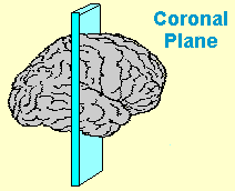


2 Note that most cases of ALS and FTD are not caused by concussions.


Read Part 1 of the series:

Brief Guide to the CTE Brains in the News. Part 1: Aaron Hernandez


References

Hobson EV, McDermott CJ. (2016). Supportive and symptomatic management of amyotrophic lateral sclerosis. Nat Rev Neurol. 12(9):526-38.

Iverson GL, Keene CD, Perry G, Castellani RJ. (2018). The Need to Separate ChronicTraumatic Encephalopathy Neuropathology from Clinical Features. J Alzheimers Dis. 61(1):17-28.

Ling H, Morris HR, Neal JW, Lees AJ, Hardy J, Holton JL, Revesz T, Williams DD. (2017). Mixed pathologies including chronic traumatic encephalopathy account fordementia in retired association football (soccer) players. Acta Neuropathol. 133(3):337-352.

Omalu B, Small GW, Bailes J, Ercoli LM, Merrill DA, Wong KP, Huang SC, Satyamurthy N, Hammers JL, Lee J, Fitzsimmons RP. (2017). Postmortem Autopsy-Confirmation of Antemortem [F-18] FDDNP-PET Scans in a Football Player With Chronic Traumatic Encephalopathy. Neurosurgery. 2017 Nov 10.


Further Reading I've written about CTE a lot, you can read more below.

FDA says no to marketing FDDNP for CTE

Is CTE Detectable in Living NFL Players?

The Ethics of Public Diagnosis Using an Unvalidated Method

The Truth About Cognitive Impairment in Retired NFL Players

Lou Gehrig Probably Died of Lou Gehrig's Disease

Blast Wave Injury and Chronic Traumatic Encephalopathy: What's the Connection?

Little Evidence for a Direct Link between PTSD and Chronic Traumatic Encephalopathy

Subscribe to Post Comments [Atom]

Monday, December 04, 2017

Brief Guide to the CTE Brains in the News. Part 1: Aaron Hernandez

Chronic traumatic encephalopathy (CTE) is the neurodegenerative disease of the moment, made famous by the violent and untimely deaths of many retired professional athletes. Repeated blows to the head sustained in contact sports such as boxing and American football can result in abnormal accumulations of tau protein (usually many years later). The autopsied brains from two of these individuals are shown below.


Left: courtesy of Dr. Ann McKee in NYT.  Right: courtesy of Dr. Bennett Omalu in CNN. These are coronal sections1 from the autopsied brains of: (L) Aaron Hernandez, aged 27; and (R) Fred McNeill, aged 63.


Both men played professional football in the NFL. Both came upon some troubled times after leaving the game. And although the CTE pathology in their brains has been attributed directly to football — repeated concussive and sub-concussive events — other potential factors have been mostly ignored. Below I'll discuss these events and phenomena, and whether they could have contributed to the condition of the post-mortem brains.


Aaron Hernandez


Illustration by Sean McCabe for Rolling Stone


Talented ex-NFL football star, PCP addict, convicted murderer, and suicide by hanging. The Rolling Stone ran two riveting articles that detailed the life (and death) of Mr. Hernandez. Despite a difficult upbringing surrounded by violence and tragedy, he was a serious and stellar athlete at Bristol High School. The tragic death of his father from a medical accident led Aaron to hang out with a less savory crowd. He fortunately ended up at the University of Florida for college football. There he failed several drug tests, but the administration mostly looked the other way. He was on a national championship team, named an all-American, and involved in a shooting where he was not charged.

Most NFL teams took a pass because of his use of recreational drugs and reputation as a hot-head:
After seeing his pre-draft psychological report, where he received the lowest possible score, one out of 10, in the category of “social maturity” and which also noted that he enjoyed “living on the edge of acceptable behavior,” a handful of teams pulled him off their boards, and 25 others let him sink like a stone on draft day.

But he ended up signing with the New England Patriots in a $40 million deal. He smoked pot constantly and avoided hanging out with the other players. “Instead of teammates, Hernandez built a cohort of thugs, bringing stone-cold gangsters over to the house to play pool, smoke chronic and carouse.” Things spiraled downwards, in terms of thug life, use of PCP (angel dust), and ultimately the murder of a friend that ended in a life sentence without parole.

He was also tried and acquitted of a separate double homicide, but his days were numbered. Two days later he hanged himself with a bedsheet in his jail cell. He was rumored to have smoked K2 (nasty synthetic cannabis) just before his death, but this was ultimately unsubstantiated.

These complicating factors lengthy history of drug abuse, death by asphyxiation must have had some effect on his brain, I mused in another post.


Meanwhile, the New York Times had a splashy piece about how the pristine brain of Aaron Hernandez presented an opportunity to study a case of “pure” CTE:
What made the brain extraordinary, for the purpose of science, was not just the extent of the damage, but its singular cause. Most brains with that kind of damage have sustained a lifetime of other problems, too, from strokes to other diseases, like Alzheimer’s. Their samples are muddled, and not everything found can be connected to one particular disease.

This was a startling statement, as I said in my secondary blog:
I’ve been struggling to write a post that highlights the misleading nature of this claim. How much of that was [the writer's] own hyperbole? Or was he merely paraphrasing the famous neuropathologists who presented their results to the media, not to peer reviewers? Is it my job to find autopsied brains from PCP abusers and suicides by hanging? Searching for the latter, by the way, will turn up some very unsavory material in forensic journals and elsewhere. At any rate, I think much of this literature glosses over any complicating elements, and neglects to mention all of the cognitively intact former football players whose brains haven’t been autopsied.

In the next post, I'll discuss the case of Fred McNeill O'Neill.


Footnote

1 Illustration of the coronal plane of section.


Read Part 2 of the series:

Brief Guide to the CTE Brains in the News. Part 2: Fred McNeill


Further Reading  
I've written about CTE a lot, you can read more below.

FDA says no to marketing FDDNP for CTE

Is CTE Detectable in Living NFL Players?

The Ethics of Public Diagnosis Using an Unvalidated Method

The Truth About Cognitive Impairment in Retired NFL Players

Lou Gehrig Probably Died of Lou Gehrig's Disease

Blast Wave Injury and Chronic Traumatic Encephalopathy: What's the Connection?

Little Evidence for a Direct Link between PTSD and Chronic Traumatic Encephalopathy


New York Times: A neuropathologist and her associate examined slices of the brain of a 27-year-old man. Credit: Boston University.

Subscribe to Post Comments [Atom]

eXTReMe Tracker