Robotto Kânibaru

Encephalon - A neuroscience carnival
Third edition available at
Thinking Meat.
Read about human-robot communication.

Robot Carnival
Subscribe to Post Comments [Atom]
Deconstructing the most sensationalistic recent findings in Human Brain Imaging, Cognitive Neuroscience, and Psychopharmacology


Subscribe to Post Comments [Atom]

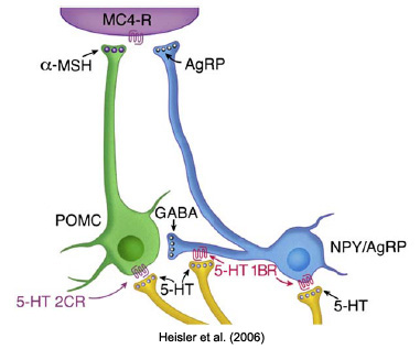
Detailed explanation [Figure 8 of Heisler et al.]: 5-HT hyperpolarizes and inhibits AgRP neurons and decreases an inhibitory drive onto POMC cells by activation of 5-HT1BRs. 5-HT also activates POMC neurons via activation of 5-HT2CRs. This leads to reciprocal increases in a-MSH release and decreases in AgRP release at MC4-R in target sites.Translated, the arcuate nucleus of the hypothalamus may play a central role in appetite suppression. The arcuate has two populatons of neurons: one expressing the anorectic melanocortin receptor agonist, a-MSH (green neuron) and the other expressing the appetitite-stimulating melanocortin receptor antagonist, AgRP (blue neuron). The combined increase in a-MSH and decrease in AgRP act at downstream MC4 receptors to suppress appetitite.
Heisler LK, Jobst EE, Sutton GM, Zhou L, Borok E, Thornton-Jones Z, Liu HY, Zigman JM, Balthasar N, Kishi T, Lee CE, Aschkenasi CJ, Zhang CY, Yu J, Boss O, Mountjoy KG, Clifton PG, Lowell BB, Friedman JM, Horvath T, Butler AA, Elmquist JK, Cowley MA. (2006). Serotonin Reciprocally Regulates Melanocortin Neurons to Modulate Food Intake. Neuron 51:239-249.However, the serotonergic drugs given to the mice in that study included D-fenfluramine, part of the scary and dangerous fen-phen fiasco. Fen-phen was linked to heart valve disease and pulmonary hypertension, and was withdrawn from the U.S. market in 1997. The mice were also given an experimental 5-HT1BR agonist not yet approved for human use.
The neural pathways through which central serotonergic systems regulate food intake and body weight remain to be fully elucidated. We report that serotonin, via action at serotonin1B receptors (5-HT(1B)Rs), modulates the endogenous release of both agonists and antagonists of the melanocortin receptors, which are a core component of the central circuitry controlling body weight homeostasis. We also show that serotonin-induced hypophagia requires downstream activation of melanocortin 4, but not melanocortin 3, receptors. These results identify a primary mechanism underlying the serotonergic regulation of energy balance and provide an example of a centrally derived signal that reciprocally regulates melanocortin receptor agonists and antagonists in a similar manner to peripheral adiposity signals.
If MC4R agonists induce spontaneous penile erections in men, this would represent a significant impediment to the development of compounds to treat obesity.
The nonselective melanocortin agonists, 1 and 3, have nausea and vomiting as adverse side effects when administered to humans either subcutaneously or intranasally. Attempts to study whether the effects of these two structurally related molecules are mechanism-based have been of limited utility.Is PT-141 really a miracle drug? That's highly doubtful. But, as PZ Myers said,
Wow. Makes me want to run out and buy stock in Palatin Technologies, the manufacturer.
Subscribe to Post Comments [Atom]
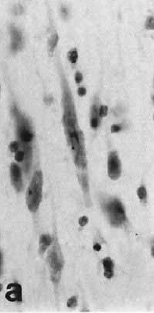
 In a neuroanatomical tour de force, Nimchinsky and colleagues (1999) obtained access to samples of the anterior cingulate cortex (and other cortical regions) from 28 different primate species, from prosimians to anthropoids to great apes to humans. They processed the samples with a Nissl stain to identify neuronal cell bodies in the cerebral cortex, a structure that (generally) consists of six layers. Spindle neurons are a unique type of neuron found in layer Vb in the ACC and frontoinsular cortex of humans. This is nothing new; spindle neurons (also called Von Economo neurons) were first identified in the 19th century by W. Betz (of the eponymous Betz cell fame, I presume) and by Nobel laureate Santiago Ramón y Cajal. What was new in 1999 was the finding that only humans and great apes have spindle neurons.
Our lab has investigated the anatomical structure of the Von Economo (spindle) neurons in anterior cingulate and fronto-insular cortex. Based on functional imaging studies of these brain areas and our studies of the expression of neurotransmitter receptors on these cells, we think they participate in fast, intuitive social decision-making. We have found that the Von Economo neurons emerge mainly in the first three years after birth. We also have evidence that in autistic subjects the Von Economo neurons are abnormally located, possibly as a result of a migration defect. This abnormality may be at least partially responsible for defective social intuition in autism.Somehow, the "spindle neuron" meme hasn't caught on like the "mirror neuron" meme. Is it because spindle neurons have been only been described anatomically (not physiologically), while the reverse is true for mirror neurons? Anatomically speaking, do we know much about mirror neurons? Here's what Rizzolatti and Craighero (2004) have to say about them:
Mirror neurons are a particular class of visuomotor neurons, originally discovered in area F5 of the monkey premotor cortex, that discharge both when the monkey does a particular action and when it observes another individual (monkey or human) doing a similar action (Di Pellegrino et al. 1992, Gallese et al. 1996, Rizzolatti et al. 1996a).In the elegantly titled article, The importance of being agranular, Stewart Shipp reviews evidence that approximately 10% of recorded cells in premotor area F5 in the macaque monkey can be classified as mirror neurons. He also points out an interesting conundrum regarding the anatomical organization of motor cortex: it's agranular, meaning it's lacking the granule cell layer (layer IV), the typical termination point for feedforward sensory information. Area 7b (or PF) in the rostral inferior parietal lobule provides the main parietal input to F5. Without going into too many details, it seems the anatomical circuitry of visual input to F5 is pretty complicated. Anyone who studies mirror neurons (or who does fMRI studies of "empathy and the mirror neuron system") should read these two papers:
from Rizzolatti G, Craighero L. (2004). The mirror-neuron system. Annu Rev Neurosci. 27:169-92.
Geyer S, Matelli M, Luppino G, Zilles K. (2000). Functional neuroanatomy of the primate isocortical motor system. Anat Embryol 202:443-74
Shipp S. (2005). The importance of being agranular: a comparative account of visual and motor cortex. Philos Trans R Soc Lond B Biol Sci. 360:797-814.
Subscribe to Post Comments [Atom]
And view the other content at KERBLOG...


a lot of people are asking me if they can post this or that drawing on their blog, or to publish it, etc.
to all the blog readers and beyond,
PLEASE NOTE THAT THERE IS NO COPYRIGHT ON ANY OF THE DRAWINGS OF THIS BLOG. PLEASE DO PUBLISH, PHOTOCOPY, PRINT, DISTRIBUTE EVERYWHERE ALL THE DRAWINGS IF YOU CAN.
when the war is over, my lawyer will call you. until then please spread them anywhere you can.
Subscribe to Post Comments [Atom]

July 23, 2006
The Haunting
By JOHN HODGMAN
New York Times Magazine
. . .
Horror, like comedy, has always been something of a reptilian-brain endeavor, unusual among the arts insofar as it is successful only when it is able to produce a single, audible emotional effect — a scream or a laugh — that is primal, cathartic and difficult to understand. This is one reason that horror has always been a director's medium: the horror movie is a contraption, and it takes a certain organizational flair to design, pace and frame a scare.

Stark R, Schienle A, Sarlo M, Palomba D, Walter B, Vaitl D. (2005). Influences of disgust sensitivity on hemodynamic responses towards a disgust-inducing film clip. Int J Psychophysiol. 57(1):61-7.
The major goal of the present functional magnetic resonance imaging study was to investigate the influence of disgust sensitivity on hemodynamic responses during disgust induction. Fifteen subjects viewed three different film excerpts (duration: 135 s each) with disgust-evoking, threatening and neutral content. The films were presented in a block design with four repetitions of each condition. Afterwards, subjects gave affective ratings for the films and answered the questionnaire for the assessment of disgust sensitivity (QADS, []). The subjects' overall disgust sensitivity was positively related to their experienced disgust, as well as to their prefrontal cortex activation during the disgust condition. Further, there was a positive correlation between subjects' scores on the QADS subscale spoilage/decay and their amygdala activation (r=0.76). This was reasonable since the disgust film clip depicted a cockroach-invasion and the subscale spoilage/decay contains, among others, an item asking for disgust towards cockroaches. The study stresses, in accordance to previous studies, the importance of considering personality traits when studying affective responses in fMRI studies.

Fundamentally, Carnival Of Souls is a visual exploration of death, which, in the course of this exploration, sees death from a subjective point of view that is at times reminiscent of Carl Dreyer's "Vampyr". However, its subjectivity is far more radical than that of the Dreyer film, since Dreyer, concentrating to some extent on actual occurences rooted in a particular time and place, dwelt only sporadically on the inner consciousness of his protagonist. By contrast, it could be argued that everything in Carnival Of Souls other than the physical death of the main character is subjective in nature. As a result, we are forced into suspension of disbelief as to the reality-status of a woman who, though materially dead, continues to experience many things through "normal" consciousness.In the study of Stark et al. (2005),
Three film clips (DISGUST, FEAR, NEUTRAL, 135 s each) with sound track were presented to the subjects. Each clip was a scene selected from a commercial movie: DISGUST showed an invasion of cockroaches, a scene from the movie Creepshow I (Romero and Rubinstein, 1982). FEAR was drawn from The Hitcher (Harmon et al., 1986) and depicted a boy threatened by a man armed with a knife. NEUTRAL came from Switzerland—The Alpine Wonderland (Scro et al., 1989) and showed urban areas.The paper was mostly about disgust and how the participants' self-ratings on a disgust sensitivity questionnaire correlated with neural responsiveness in the orbitofrontal cortex, medial prefrontal cortex and the amygdala. Are the contestants on Fear [and Disgust] Factor screened with such questionnaires before going on the show??

Subscribe to Post Comments [Atom]
Now back to our irregularly scheduled neuroscience programming!

an online database of published functional neuroimaging experiments with coordinate-based (Talairach) activation locations. The goal of BrainMap is to provide a vehicle to share methods and results of brain functional imaging studies. It is a tool to rapidly retrieve and understand studies in specific research domains, such as language, memory, attention, reasoning, emotion, and perception, and to perform meta-analyses of like studies.where one can do
coordinate-based meta-analyses according to the activation likelihood estimation (ALE) method.That's when I did the ALE meta-analysis for imaging studies of physical pain, for comparison to "empathy for pain" and the "social rejection of being excluded from Pong" (which, of course, feels exactly like being cut open with a scalpel).
Eisenberger NI, Lieberman MD, Williams KD. (2003). Does rejection hurt? An FMRI study of social exclusion. Science 302: 290-2.More on that topic later.
A neuroimaging study examined the neural correlates of social exclusion and tested the hypothesis that the brain bases of social pain are similar to those of physical pain. Participants were scanned while playing a virtual ball-tossing game in which they were ultimately excluded. Paralleling results from physical pain studies, the anterior cingulate cortex (ACC) was more active during exclusion than during inclusion and correlated positively with self-reported distress. Right ventral prefrontal cortex (RVPFC) was active during exclusion and correlated negatively with self-reported distress. ACC changes mediated the RVPFC-distress correlation, suggesting that RVPFC regulates the distress of social exclusion by disrupting ACC activity.
Somerville LH, Heatherton TF, Kelley WM. (2006). Anterior cingulate cortex responds differentially to expectancy violation and social rejection. Nat Neurosci. Jul 2 [Epub ahead of print]Now back to physical pain. OK, here's another ALE map that shows clusters of activation (below). In this map, different clusters of activation are coded with different colors. The first map (above) was color-coded for significance level.
This study investigated human anterior cingulate cortex (ACC) involvement during a task that dissociated expectancy violation from social rejection. Across two studies, participants underwent functional magnetic resonance imaging while making social judgments and receiving fictitious feedback that was either positive or negative and consistent or inconsistent with their expectations. The results demonstrate that the dorsal ACC is sensitive to expectancy violations, whereas the ventral ACC is differentially responsive to social feedback.]

Meurisse M, Defechereux T, Hamoir E, Maweja S, Marchettini P, Gollogly L, Degauque C, Joris J, Faymonville ME. (1999). Hypnosis with conscious sedation instead of general anaesthesia? Applications in cervical endocrine surgery. Acta Chir Belg. 99:151-8.Uh, NO!
Subscribe to Post Comments [Atom]
Sometimes, Cognitive Neuroscience just doesn't seem very important.
STARRY NIGHT (excerpt) 6.31 min
a minimalistic improvisation by:
mazen kerbaj / trumpet
the israeli air force / bombs
recorded by mazen kerbaj on the balcony of his flat in beirut,
on the night of 15th to 16th of july 2006.
© mazen kerbaj 2006
Subscribe to Post Comments [Atom]
A metaphor is a glorious thing
A diamond ring
The first day of summer
A metaphor is a breath of fresh air
A turn-on
An aphrodisiac
--Sparks
"Metaphor" <----- CLICK HERE
[OPEN IN NEW TAB OR WINDOW AND LISTEN WHILE READING]
Alternate perspectives from Cognitive Linguistics can be found here:
Don't don't don't don't
Don't mix them
We we we
Won't mix them
--ibid
Chicks dig dig
d-i-g
dig dig metaphors
--ibid
Use them wisely
Use them well
And you'll never know the hell
Of loneliness
--ibid
Subscribe to Post Comments [Atom]
Subscribe to Post Comments [Atom]
in a carnival
of sights to see
all the cheap thrill seekers
the vendors and the dealers
they crowded around me
"Carnival"
-Natalie Merchant
have I been blind
have I been lost
inside my self and
my own mind
hypnotized
mesmerized
by what my eyes have seen?
-ibid
Subscribe to Post Comments [Atom]
 You've probably heard about the case of Terry Wallis, a 42 year old man in Arkansas who spent 19 years in a "minimally conscious state" following severe traumatic brain injury. Then one day he saw his mother walk into the room and (after not speaking all those years) said "Mom."
The Aspen workgroup defined the minimally conscious state (MCS) as a condition of severely altered consciousness in which the person demonstrates minimal but definite behavioral evidence of self or environmental awareness (Giacino et al, 1997).A number of bloggers have commented on how his case is different from that of Terry Schiavo, who was in a persistent vegetative state (not a MCS).
Using a novel technique, they saw evidence of new growth in the midline cerebellum, an area involved in motor control, as Mr. Wallis gained strength and range in his limbs. Another area of new growth, located along the back of the brain, is believed by some experts to be a central switching center for conscious awareness.The "central switching center for conscious awareness" is the precuneus, interestingly enough.
The daily exercises, the interactions with his parents, his regular dose of antidepressant medication: any or all of these might have spurred brain cells to grow more connections, the researchers said.
"The big missed opportunity is that we didn't know this guy would spontaneously emerge, and we didn't get to monitor him before then" to find out what preceded it, Dr. Schiff said.
Henning U. Voss, Aziz M. Uluç, Jonathan P. Dyke, Richard Watts, Erik J. Kobylarz, Bruce D. McCandliss, Linda A. Heier, Bradley J. Beattie, Klaus A. Hamacher, Shankar Vallabhajosula, Stanley J. Goldsmith, Douglas Ballon, Joseph T. Giacino and Nicholas D. Schiff. (2006). Possible axonal regrowth in late recovery from the minimally conscious state. J. Clin. Invest. 116: 2005-2011. OPEN ACCESS ARTICLE!In a commentary on the article, Laureys, Boly, and Maquet note the importance of the precuneus in conscious awareness:
We used diffusion tensor imaging (DTI) to study 2 patients with traumatic brain injury. The first patient recovered reliable expressive language after 19 years in a minimally conscious state (MCS); the second had remained in MCS for 6 years. Comparison of white matter integrity in the patients and 20 normal subjects using histograms of apparent diffusion constants and diffusion anisotropy identified widespread altered diffusivity and decreased anisotropy in the damaged white matter. These findings remained unchanged over an 18-month interval between 2 studies in the first patient. In addition, in this patient, we identified large, bilateral regions of posterior white matter with significantly increased anisotropy that reduced over 18 months. In contrast, notable increases in anisotropy within the midline cerebellar white matter in the second study correlated with marked clinical improvements in motor functions. This finding was further correlated with an increase in resting metabolism measured by PET in this subregion. Aberrant white matter structures were evident in the second patient’s DTI images but were not clinically correlated. We propose that axonal regrowth may underlie these findings and provide a biological mechanism for late recovery. Our results are discussed in the context of recent experimental studies that support this inference.
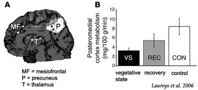
The most remarkable finding in the Voss et al. study (12) was the MRI assessment of transiently increased fractional anisotropy and directionality in the posterior midline cortices (encompassing the cuneus and precuneus), interpreted as increased myelinated fiber densities and novel corticocortical sprouting, paralleling the emergence of the patient from MCS. The same area of the patient’s brain also showed amplified metabolic activity, as measured by PET. This finding stresses the importance of the posterior medial structures in consciousness of self and interaction with the environment (14, 15). Activity in the medial parietal cortex (i.e., precuneus) seems to show it to be the brain region that best differentiates MCS from VS patients (16). Interestingly, this area is among the most active brain regions in conscious waking (15) and is among the least active in altered states of consciousness, such as pharmacological coma (17), sleep (18), dementia (19), Wernicke-Korsakoff syndrome, and postanoxic amnesia (20). It has been suggested that this richly connected multimodal posteromedial associative area is part of the neural network subserving human awareness (21).
 The figure above illustrates Mr. Wallis' brain. On the left is a DTI scan to trace fiber tracts. On the right is a PET scan to measure glucose metabolism. The areas highlighted are on the posterior medial surface of the brain, the parietal-occipital region, which includes the cuneus and precuneus.
Steven Laureys, Mélanie Boly and Pierre Maquet (2006). Tracking the recovery of consciousness from coma. J. Clin. Invest. 116:1823-1825. OPEN ACCESS ARTICLE!
Predicting the chances of recovery of consciousness and communication in patients who survive their coma but transit in a vegetative state or minimally conscious state (MCS) remains a major challenge for their medical caregivers. Very few studies have examined the slow neuronal changes underlying functional recovery of consciousness from severe chronic brain damage. A case study in this issue of the JCI reports an extraordinary recovery of functional verbal communication and motor function in a patient who remained in MCS for 19 years (see the related article beginning on page 2005). Diffusion tensor MRI showed increased fractional anisotropy (assumed to reflect myelinated fiber density) in posteromedial cortices, encompassing cuneus and precuneus. These same areas showed increased glucose metabolism as studied by PET scanning, likely reflecting the neuronal regrowth paralleling the patient’s clinical recovery. This case shows that old dogmas need to be oppugned, as recovery with meaningful reduction in disability continued in this case for nearly 2 decades after extremely severe traumatic brain injury.
Mute 19 Years, He Helps Reveal Brain's Mysteries
. . .
He does not feel any physical pain, he told his parents, and he has no real sense of time. He also said recently that he was "proud" to be alive.
"It is good to know all that," said his father, sitting on the porch on Saturday evening.
"It's good to hear him say that, because if he didn't say so, you'd just have no way to know."
Subscribe to Post Comments [Atom]
 Encephalon - A neuroscience carnival
Subscribe to Post Comments [Atom]
Subscribe to Post Comments [Atom]

I don't do crack.
cuz crack is cheap
crack is cheap, OK
crack is wack!
I do freebase now.

Subscribe to Post Comments [Atom]