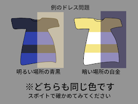
もう何番煎じかも分からないけど例のドレス問題をまとめてみました。青黒/白金に見える人の色覚やモニタを疑ってる人はぜひご覧ください。 pic.twitter.com/6euNYw9xUa
— ぶどう茶 (@budoucha) February 27, 2015
Could one's chronotype (degree of "morningness" vs. "eveningness") be related to your membership on Team white/gold vs. Team blue/black?
Dreaded by night owls everywhere, Daylight Savings Time forces us to get up an hour earlier. Yes, [my time to blog and] I have been living under a rock, but this evil event and an old tweet by Vaughan Bell piqued my interest in melanopsin and intrinsically photosensitive retinal ganglion cells.
Totally speculative: wonder whether perceptual diffs reflect diffs in melanopsin. Blue sensitive, mediates brightness http://t.co/841bN6zvCs
— Vaughan Bell (@vaughanbell) February 28, 2015
I thought this was a brilliant idea, perhaps differences in melanopsin genes could contribute to differences in brightness perception. More about that in a moment.
{Everyone already knows about #thedress from Tumblr and Buzzfeed and Twitter obviously}
In the initial BuzzFeed poll, 75% saw it as white and gold, rather than the actual colors of blue and black. Facebook's more systematic research estimated this number was only 58% (and influenced by probably exposure to articles that used Photoshop). Facebook also reported differences by sex (males more b/b), age (youngsters more b/b), and interface (more b/b on computer vs. iPhone and Android).
Dr. Cedar Riener wrote two informative posts about why people might perceive the colors differently, but Dr. Bell was not satisfied with this and other explanations. Wired consulted two experts in color vision:
“Our visual system is supposed to throw away information about the illuminant and extract information about the actual reflectance,” says Jay Neitz, a neuroscientist at the University of Washington. “But I’ve studied individual differences in color vision for 30 years, and this is one of the biggest individual differences I’ve ever seen.”and
“What’s happening here is your visual system is looking at this thing, and you’re trying to discount the chromatic bias of the daylight axis,” says Bevil Conway, a neuroscientist who studies color and vision at Wellesley College. “So people either discount the blue side, in which case they end up seeing white and gold, or discount the gold side, in which case they end up with blue and black.”
Finally, Dr. Conway threw out the chronotype card:
So when context varies, so will people’s visual perception. “Most people will see the blue on the white background as blue,” Conway says. “But on the black background some might see it as white.” He even speculated, perhaps jokingly, that the white-gold prejudice favors the idea of seeing the dress under strong daylight. “I bet night owls are more likely to see it as blue-black,” Conway says.
Melanopsin and Intrinsically Photosensitive Retinal Ganglion Cells
Rods and cones are the primary photoreceptors in the retina that convert light into electrical signals. The role of the third type of photoreceptor is very different. Intrinsically photosensitive retinal ganglion cells (ipRGCs) sense light without vision and:
- ...play a major role in synchronizing circadian rhythms to the 24-hour light/dark cycle [via direct projections to the suprachiasmatic nucleus]...
- ...contribute to the regulation of pupil size and other behavioral responses to ambient lighting conditions...
- ...contribute to photic regulation of, and acute photic suppression of, release of the hormone melatonin...
Recent research suggests that ipRGCs may play more of a role in visual perception than was originally believed. As Vaughan said, melanopsin (the photopigment in ipRGCs) is involved in brightness discrimination and is most sensitive to blue light. Brown et al. (2012) found that melanopsin knockout mice showed a change in spectral sensitivity that affected brightness discrimination; the KO mice needed higher green radiance to perform the task as well as the control mice.
The figure below shows the spectra of human cone cells most sensitive to Short (S), Medium (M), and Long (L) wavelengths.
Spectral sensitivities of human cone cells, S, M, and L types. X-axis is in nm.
The peak spectral sensitivity for melanopsin photoreceptors is in the blue range. How do you isolate the role of melanopsin in humans? Brown et al. (2012) used metamers, which are...
...light stimuli that appear indistinguishable to cones (and therefore have the same color and photopic luminance) despite having different spectral power distributions. ... to maximize the melanopic excitation achievable with the metamer approach, we aimed to circumvent rod-based responses by working at background light levels sufficiently bright to saturate rods.
They verified their approach in mice, then used a four LED system to generate stimuli that diffed in presumed melanopsin excitation, but not S, M, or L cone excitation. All six of the human participants perceived greater brightness as melanopsin excitation increased (see Fig. 3E below). Also notice the individual differences in test radiance with the fixed 11% melanopic excitation (on the right of the graph).
Modified from Fig. 3E (Brown et al. (2012). Across six subjects, there was a strong correlation between the test radiance at equal brightness and the melanopic excitation of the reference stimulus (p < 0.001).1
Maybe Team white/gold and Team blue/black differ on this dimension? And while we're at it, is variation in melanopsin related to circadian rhythms, chronotype, even seasonal affective disorder (SAD)? 2 There is some evidence in favor of the circadian connections. Variants of the melanopsin (Opn4) gene might be related to chronotype and to SAD, which is much more common in women. Another Opn4 polymorphism may be related to pupillary light responses, which would affect light and dark adaptation. These genetic findings should be interpreted with caution, however, until replicated in larger populations.
Could This Device Hold the Key to “The Dress”?
ADDENDUM (March 10 2015): NO, according to Dr. Geoffry K. Aguirre of U. Penn.: “Speaking as a guy with a 56-primary version of This Device to study melanopsin, I think the answer to your question is 'no'…” His PNAS paper, Opponent melanopsin and S-cone signals in the human pupillary light response, is freely available.3
A recent method developed by Cao, Nicandro and Barrionuevo (2015) increases the precision of isolating ipRGC function in humans. The four-primary photostimulator used by Brown et al. (2012) assumed that the rod cells were saturated at the light levels they used. However, Cao et al. (2015) warn that “a four-primary method is not sufficient when rods are functioning together with melanopsin and cones.” So they:
...introduced a new LED-based five-primary photostimulating method that can independently control the excitation of melanopsin-containing ipRGC, rod and cone photoreceptors at constant background photoreceptor excitation levels.
Fig. 2 (Cao et al., 2015). The optical layout and picture of the five-primary photostimulator.
Their Journal of Vision article is freely available, so you can read all about the methods and experimental results there (i.e., I'm not even going to try to summarize them here).
So the question remains: beyond the many perceptual influences that everyone has already discussed at length (e.g., color constancy, Bayesian priors, context, chromatic bias, etc.), could variation in ipRGC responses influence how you see “The Dress”?
Footnotes
1Fig 3E (continued). The effect was unrelated to any impact of melanopsin on pupil size. Subjects were asked to judge the relative brightness of three metameric stimuli (melanopic contrast −11%, 0%, and +11%) with respect to test stimuli whose spectral composition was invariant (and equivalent to the melanopsin 0% stimulus) but whose radiance changed between trials.
2 This would test Conway's quip that night owls are more likely to see the dress as blue and black.
3 Aguirre also said that a contribution from melanopsin (to the dress effect) was doubtful, at least from any phasic effect: “It's a slow signal with poor spatial resolution and subtle perceptual effects.” It remains to be seen whether any bias towards discarding blue vs. yellow illuminant information is affected by chronotype.
Interesting result from Spitschan, Jain, Brainard, & Aguirre 2014):
The opposition of the S cones is revealed in a seemingly paradoxical dilation of the pupil to greater S-cone photon capture. This surprising result is explained by the neurophysiological properties of ipRGCs found in animal studies.
References
Brown, T., Tsujimura, S., Allen, A., Wynne, J., Bedford, R., Vickery, G., Vugler, A., & Lucas, R. (2012). Melanopsin-Based Brightness Discrimination in Mice and Humans. Current Biology, 22 (12), 1134-1141 DOI: 10.1016/j.cub.2012.04.039
Cao, D., Nicandro, N., & Barrionuevo, P. (2015). A five-primary photostimulator suitable for studying intrinsically photosensitive retinal ganglion cell functions in humans. Journal of Vision, 15 (1), 27-27 DOI: 10.1167/15.1.27


I think there something to be said for the idea of assumptions about the illuminant being different between those seeing one versus the other color pair. I read something on the SfN site that had a nice figure showing the dress with a common color constancy illusion as background, and I could see the two variations for the first time. I forget where that was though. I'm the worst night owl you'll ever find but could not see the black and blue thing.
ReplyDeleteFound the link - the cool jpeg is near the bottom. http://blog.brainfacts.org/2015/03/what-color-is-distress/#.VP5yArPF-iZ
ReplyDeleteThanks for the link, I hadn't seen that article. The cropped version of the image by Rosa Lafer-Sousa is even more striking. I agree with you about the illuminant, but what biases people to discount blue vs. yellow?
ReplyDeleteI know I'm way late to the party (too many other major life events), but I only see blue and black (and the top of the dress has always seemed more brown to me)...
Your thoughtful blog post deserves a more complete response than the one tweet I offered.
ReplyDeleteWith my collaborators Manuel Spitschan and David Brainard, we have been studying the effects of melanopsin stimulation in humans, using a 56-primary version of the device described by Cao and colleagues. All those extra primaries come in handy for creating stimulus modulations that are very precisely targeted at melanopsin, with minimal stimulation of the cones. The extra primaries are used to cover the imperfect specification of cone spectral sensitivity, preceptoral filtering, and even the possibility of cones positioned under retinal blood vessel responding to stimulation.
We have been looking at the perceptual effects of melanopsin stimulation. My first response to your proposal (that individual differences in melanopsin function contribute to differences in The Dress perception) was "no way". In my tweet, I offered some reasons for this impression, namely that melanopsin is sluggish in its responses and has very poor spatial resolution. Upon further reflection, I think these properties of melanopsin do not actually speak against your hypothesis. In fact, a slowly changing signal that integrates over the entire image is not a bad candidate for this individual difference!
A better reason to be skeptical of a role for melanopsin is that the varying perception of the dress is linked tightly to color constancy (which, I hasten to add, is not my area of expertise). Placing the image in a blue or yellow background is sufficient to change the percept for most observers. This is a chromatic mechanism at work.
It would be a surprise to discover that melanopsin contributes to interpretation of the chromatic content of the illuminant, but we are still in the early stages of understanding the role of this photopigment. Current results suggest that it contributes to an achromatic mechanism of brightness perception. Interestingly, however, we find that there is a fair bit of individual difference in this effect upon brightness perception. Motivated by your blog post, I'll start having subjects give us their interpretation of the dress image, and see if anything surprising turns up!
Thanks for the informative update, Geoff. Will await the results of your current study!
ReplyDelete