Monday, December 30, 2013

How Can We Forget?

** This post is meant to be read in tandem with its more complimentary cousin, Electroconvulsive Therapy Impairs Memory Reconsolidation, at The Neurocomplimenter. **


spECTrum 5000Q® ECT device (MECTA)


Bad memories haunt a significant number of people with serious mental illnesses, such as chronic major depression and post-traumatic stress disorder (PTSD). If it were possible to undergo an experimental procedure that selectively impairs your memory for an extremely unpleasant event, would you do it? If this sounds like the plot of Eternal Sunshine of the Spotless Mind, you're not alone.

A pet peeve of mine is reference to this excellent but far-fetched film in scientific journals and popular media coverage of “memory erasure.” The idea that it's possible to selectively remove a complex autobiographical memory that has become intimately entwined with the fabric of our constructed selves is utter science fiction.


At some level, even Michel Gondry knew it. One incident in Eternal Sunshine is suggestive of how memories might actually be stored. It was after one of the main characters (Joel) had his memories of his ex-girlfriend Clementine erased, and he couldn't remember who Huckleberry Hound was. He had associated the cartoon character and the song "Darling Clementine" with her. That resembles a semantic network, where an overlapping network of neurons and synapses code different but semantically related things. Take out all episodic and semantic memories of Clementine, and knowledge of Huckleberry Hound goes with it.

The latest incarnation of this particular memory erasure meme was provoked by publication of a paper (Kroes et al., 2013) that examined the process of memory reconsolidation in depressed patients administered a course of electroconvulsive therapy (ECT). Here are some of the headlines:
Zapping the brain can help to spot-clean nasty memories

Absolutely shocking: electrocuting brain can wipe unpleasant memories

Unwanted Memories Erased in Electroconvulsive Therapy Experiment

Shocking Memories Away

My companion post at The Neurocomplimenter reviews the literature on memory reconsolidation and describes the experiment of Kroes et al. (2013) in some detail. What I'd like to do here is to point out possible weaknesses in the results that could undermine the authors' conclusions. I'll also discuss a much earlier ECT study which did not support the notion that reactivated memories are especially vulnerable to disruption (Squire et al., 1976).

To briefly reiterate the methods used in the new paper (Kroes et al., 2013), the participants were 39 patients with moderate to severe major depression. They were either at the end of an acute treatment cycle or receiving maintenance ECT. The study used a between-subjects design with three different experimental conditions, with patients randomly assigned to one of the three groups (n=13 in each). The within-subjects factor was whether or not the patients received a reminder of previously learned material before treatment.

All participants learned two different emotionally charged slide stories with audio narration, each consisting of 11 images. In one, a boy is in an accident that severs his feet, which are reattached at the hospital. In the other, two sisters leave their home at night, and one is kidnapped at knife point and attacked by an escaped convict.

Memory for one of the stories was reactivated a week later by presenting part of the first slide, and then giving a test for this slide. Only four minutes later, Groups A and B were anesthetized and received ECT. Group C received their ECT treatment at a later date. The final memory test for Groups A and C was 24 hrs after the reminder, while Group B was tested as soon as they woke up from the procedure (mean = 104 min later). The final test consisted of 40 multiple choice questions about each of the stories.

The basic idea is that reconsolidation of the reactivated story isn't complete within 104 min, so Group B's test performance should be the same for the two stories. In contrast, reconsolidation is complete by 24 hrs, so for Group A the disruptive effect of ECT should selectively impair memory for the transiently reactivated story, which is in a labile state (relative to the "consolidated" story learned 7 days earlier).

Is that what was observed? Statistically speaking, yes. But two patients in Group B (out of 13 total) performed very well on the reactivated story (see purple box in the figure below).


Fig. 1 (modified from Kroes et al. 2013). ECT disrupts reconsolidation. Memory scores on the multiple choice test are expressed as percentage correct (y axis). Memory for the reactivated story shown in solid bars and non-reactivated story in open bars. Each circle is the score for an individual patient. The horizontal dotted line is chance performance. Group A is in red, Group B in blue, and Group C in orange. Edited to add: The purple box highlights 2 outliers in Group B who could be driving the major effect.


If these two individuals are omitted, would the difference between Groups A and B still be significant? This is the key finding of the paper, that memory for the reactivated story is no better than chance if ECT disrupts reconsolidation (a time-dependent process). Hence all the Eternal Sunshine / “memory erasure” headlines.


The purple boxes show that (1) the Reactivation x Group interaction squeaks in at just barely significant (p=.049); and (2) the Group A vs. Group B comparison for the reactivated story is p=.042. Clearly, it would be nice to include twice as many patients in each group. But it took the authors 3.5 years to recruit their final total of n=39.

This type of study is not easy to pull off, which is why I applaud the authors (and the patients) for such an ambitious undertaking. I thought it was a very clever idea as well, but not an original one as it turns out.

In the 1970s and 80s, Dr. Larry Squire and his colleagues published a series of papers on ECT and memory. The one I'll describe here takes a similar approach to Kroes et al. by testing previously learned material after ECT, and by giving a memory reminder just before the treatment (Squire et al., 1976).

Squire et al. (1976) used a completely within-subjects design (n=12) that manipulated the pre-ECT learning interval (14-18 hrs vs. 3-10 min). The third condition presented a memory reminder 3-10 min before ECT for material learned 14-18 hrs previously. Completely different stimuli were used each time, and the order of conditions was counterbalanced. The memory tests were recognition memory for a set of 32 previously learned items (common objects, common words, yearbook photos, and nonsense drawings), and paired associates (producing the correct target for 18 previously learned cue-target pairs). In all conditions, retention was tested 6-10 hrs after ECT (compare to 104 min and 24 hrs in Kroes et al.).

A separate group of patients (n=9) was tested on their remote memories for old TV shows under three conditions: (1) 6-10 hrs after ECT; (2) 14-18 hrs before  ECT and again with the same questions 6-10 hr after ECT; (3) Less than 10 min before ECT and again with the same questions 6-10 hrs after ECT (the reminder procedure).

The critical result is that the memory reactivation procedure did not impair performance (bar graphs A vs. R below). In both of these conditions, material was learned 14-18 hrs before ECT. This is in contrast to the findings of Kroes et al. (2013).


Fig. 1 (modified from Squire et al., 1976). Results from (A) 32-item recognition memory test, and (B) paired-associate learning test, under three conditions in conjunction with the patients' first 3 ECT sessions. Retention was significantly impaired in Condition B (initial learning 3-10 min before ECT). The reminder procedure (Condition R) caused no impairment in performance relative to Condition A.


Squire and colleagues (1976) concluded that “...the results provide no evidence that the presentation of previously learned material just prior to ECT increases its vulnerability to disruption.” Similar results were observed in the patient group tested on their knowledge of old TV shows: “the results clearly indicate that amnesia for remote memory did not occur when remote memory was evoked prior to ECT.”

The final conclusions [clairvoyantly] throw cold water on the study published 37 years later:
The present findings also have important clinical implications. The reactivation phenomenon described in experimental animals has raised the possibility that it might be therapeutically advantageous to evoke depressive ideation just prior to treatment, in order to produce amnesia for this ideation. The results reported here strongly suggest that this procedure would be ineffective.

However, you'll probably notice some differences between the two studies. Kroes et al. pointed out that their effect was observed at a 24 hr retention interval, while Squire et al. only tested at 6-10 hrs (perhaps not long enough to disrupt reconsolidation). The Squire stimuli were neutral in valence, whereas the Kroes stimuli were emotional (and perhaps more susceptible to disruption). There were also differences in the patient groups (Squire's were younger, mean=39 yrs), anesthesia used, electrode locations, and ECT parameters (likely to be way more potent in the earlier study, which would predict worse amnesia). An unfortunate side effect of ECT is memory impairment, although other studies claim the opposite.

Certainly, subjective cognitive complaints after ECT are very common. For a first hand look at some of the more devastating effects, watch the powerful video below. For a lighthearted and critically acclaimed look at fictional memory erasure, watch Eternal Sunshine of the Spotless Mind.


References

Kroes MC, Tendolkar I, van Wingen GA, van Waarde JA, Strange BA, & Fernández G (2013). An electroconvulsive therapy procedure impairs reconsolidation of episodic memories in humans. Nature neuroscience PMID: 24362759

Squire LR, Slater PC, & Chace PM (1976). Reactivation of recent or remote memory before electroconvulsive therapy does not produce retrograde amnesia. Behavioral biology, 18 (3), 335-43 PMID: 1016174


Liz Spikol (of The Trouble With Spikol fame) tries to explain the confusion and the loss of self she felt after waking up from ECT.


"After the ECT, I did not know how to use a toothbrush. And that lasted for months."
- Liz Spikol (at 9:28)

Subscribe to Post Comments [Atom]

Friday, December 27, 2013

The Creativity of Denial


Never Forget Srebrenica, by Scott McIntyre. A Bosnian Muslim man makes his way past the caskets of those killed in the Srebrenica genocide of July 1995.


Horrible, unspeakable memories will forever haunt the psyches of many survivors of war, genocide, and other atrocities. But what is behind the systematic denial of crimes against humanity?

The Science of Hatred

What makes humans capable of horrific violence? Why do we deny atrocities in the face of overwhelming evidence? A small group of psychologists say they are moving toward answers. Is anyone listening?

By Tom Bartlett

The former battery factory on the outskirts of Srebrenica, a small town in eastern Bosnia, has become a grim tourist attraction. Vans full of sightseers, mostly from other countries, arrive here daily to see the crumbling industrial structure, which once served as a makeshift United Nations outpost and temporary haven for Muslims under assault by Serb forces determined to seize the town and round up its residents. In July 1995 more than 8,000 Muslim men, from teenagers to the elderly, were murdered in and around Srebrenica, lined up behind houses, gunned down in soccer fields, hunted through the forest.

This stunning article in The Chronicle of Higher Education focuses on studies of intergroup conflict, in particular the work of Sabina Cehajic-Clancy, a Bosnian social psychologist. “It is unbelievable the extent and amount of creativity that people possess when it comes to denying,” she said.

Sadly, this sort of research is seriously undervalued in psychology:
Studying conflict can be a draining, thankless endeavor. Government officials rarely turn to social psychologists for advice on how to end war or cool simmering tensions. Within psychology, research on intergroup conflict is not a speedy route to professional acclaim. The fieldwork can be arduous and expensive. Funds are hard to come by, and so is publication in top journals. You’d be better off surveying undergraduates about their dating preferences or dietary habits.

Subscribe to Post Comments [Atom]

Saturday, December 21, 2013

When Waking Up Becomes the Nightmare: Hypnopompic Hallucinatory Pain


Most of us have had frightening nightmares – someone is chasing after us trying to kill us, or the world is coming to an end. Other disturbing dreams are based on real life anxieties – our partner leaves us, we lose our job, we become homeless. One specific psychiatric condition includes nightmares as part of the diagnosis. Individuals with post-traumatic stress disorder (PTSD) often have terrible nightmares that relive the traumatic event (Pigeon et al., 2013)

We're always glad to wake up from such nightmares, whether they were of the supernatural or mundane or terrifying variety. "Thank god it was only a dream," we say.

But what if waking up from sleep was the nightmare? Hypnopompic hallucinations are unusual sensory phenomena experienced just before or during awakening. Their better known mirror image, hypnagogic hallucinations, are vivid and frightening episodes of seeing or hearing or feeling phantom sensations while falling asleep (or in early stage 1 sleep). Both are frequently associated with sleep paralysis, the terrifying condition of being half awake but unable to move. This is because the complete muscle atonia typically experienced during REM sleep has oozed into lighter stages of non-REM sleep.


Hypnagogic and hypnopompic hallucinations are usually associated with narcolepsy, but 37% of a representative community sample reported frequent hypnagogic hallucinations, and 12.5% reported hypnopompic hallucinations (Ohayon et al., 1996).1 This went well beyond the low incidence of narcolepsy in that population. Both types of hallucinations were more common in those with insomnia, excessive daytime sleepiness, anxiety disorders, and depression (according to self-report).


Night Terrors 1, by Beth Robinson


Nocturnal Episodes of Pain and Screaming

A new case study in the journal Sleep (Mantoan et al., 2013) reports on the terrifying hypnopompic hallucinations of a 43 year old woman who experiences intense limb pain when waking up, which vanishes within 30 seconds. This is a very unusual manifestation of a non-REM parasomnia, a sleep disorder involving partial arousal during the transition between non-REM and wakefulness. The phenomenology might be best characterized as a night terror.

According to the case report (Mantoan et al., 2013), the patient had...
...a history of nocturnal screaming episodes within 1–2 h of sleep onset from the age of 30 years. Her husband was habitually awoken by his wife screaming loudly, usually flapping either her right or left hand against the bed in a semi-purposeful fashion. Her husband reported that the events were sometimes heralded by an inspiratory sigh, she looked terrified and would not respond to him. The screaming would usually last 5–10 sec, and she would then complain to her husband of intense pain affecting the fingers of either hand or arm and occasionally her legs, with no associated numbness or paraesthesia. She would become fully orientated within 30 sec and would be partially amnesic for the event, but would recall an accompanying sense of “fighting to stay alive” associated with intense panic and often accompanied by fast regular palpitations. Otherwise no dream mentation or visualizations were reported in association with the episodes.

She initially had these episodes monthly, but they increased in frequency to 2-5 times a week with 1-2 episodes per night.
She was unable to identify any triggers for the episodes, and neither she nor her husband considered her to be stressed, anxious, or depressed. There was no history of sleep violence, sleep sex, sleep eating, or any other NREM parasomniac automatisms. 

The authors could not identify any standard physical source for the pain. Thoracic outlet syndrome, cervical radiculopathy, focal nerve entrapment, and median neuropathy (carpal tunnel syndrome) were all ruled out.

Pharmacological treatments were unsuccessful. A low dose (0.5–1 mg) of clonazepam was poorly tolerated (it made her feel depressed) and had no effect on her symptoms. Paroxetine was poorly tolerated (due to sedative effects), and gabapentin was also a complete failure. Trazodone, a sedating antidepressant most often prescribed for insomnia, actually made the symptoms worse.

An MRI ruled out a thalamic or hypothalamic lesion. Sleep EEG revealed sudden arousals from deep sleep, accompanied by looks of pain and/or fear on the patient's face. The episodes were consistent with a NREM parasomnia. In the example below, the patient was shaking her arm – muscle activity (EMG) is shown in the green trace.


adapted from Fig. 1B (Mantoan et al., 2013). EEG showing delta waves of stage 3 sleep before an episode of arousal with shaking of one arm and looks of fear. Channels 1-12 are EEG; channels 13 and 14 are electro-oculogram (EOG) activity; channel 15 is electromyography (EMG); channel 16 is electrocardiogram (ECG); channel 17 is oxygen saturation by pulse oximetry (SpO2).  {click on image for a larger view}


What did the doctors do to help this poor woman? Nothing, it seems. A few more musculoskeletal causes need to be ruled out.

The authors end on a vague note about the possible mechanism(s):
In conclusion, to our knowledge this is the first report of a NREM parasomnia associated with painful paroxysms, for which we postulate the following underlying pathophysiological mechanism: an internal or external stimulus triggers arousal, facilitating the activation of innate motor pattern generators in the brainstem and activating somatosensory cortical areas to produce hypnopompic hallucinatory pain.

So instead of the more typical visual hallucinations, the patient experiences pain hallucinations that originate.... where?? It seems to me that the sleep EEG could be analyzed more thoroughly, beyond merely ruling out seizure occurrence. Perhaps another imaging modality like PET could be tried (PET would be quieter than fMRI and would better tolerate movement). Identifying the neurophysiological correlates of her phantom night terror pain would provide a fascinating glimpse into a highly unusual sensory phenomenon.2


Further Reading

The Phenomenology of Pain During REM Sleep

The Neurophysiology of Pain During REM Sleep


Footnotes

1 The questions asked in the telephone interviews by Ohayon et al. (1996) were:
(a) Do you experience at least twice a week the following perceptions?

(i) the realistic feeling that someone or something is present in the room
(ii) a vivid experience of being caught in a fire
(iii) a vivid experience that you are about to be attacked
(iv) a vivid experience that you are flying through the air
(v) the feeling that you will soon fall into an abyss
(vi) the feeling that shadows or objects are moving and distorting.

(b) Do you experience other types of vivid perceptions?

(c) Can you specify the type of perception?

(i) auditory
(ii) visual
(iii) kinetic (involving movement)
To me, the most surprising part of the survey is that 37% reported these phenomenon at sleep onset twice a week for the past year. This contrasts sharply with only 0.04% reporting symptoms of narcolepsy.

2 I've occasionally felt pain in dreams that vanished upon awakening, but I'm pretty sure the episodes occurred during REM (or another stage of dreaming sleep), because visual narrative content was associated with the episodes. Those experiences were clearly not night terrors, and very different from what was reported in the case study.


References

Mantoan L, Eriksson SH, Nisbet AP, & Walker MC (2013). Adult-onset NREM parasomnia with hypnopompic hallucinatory pain: a case report. Sleep, 36 (2), 287-90 PMID: 23372277

Ohayon MM, Priest RG, Caulet M, & Guilleminault C (1996). Hypnagogic and hypnopompic hallucinations: pathological phenomena? The British journal of psychiatry, 169 (4), 459-67 PMID: 8894197

Pigeon WR, Campbell CE, Possemato K, Ouimette P. (2013). Longitudinal relationships of insomnia, nightmares, and PTSD severity in recent combat veterans. J Psychosom Res. 75:546-50.


The Sleep Paralysis Project

Subscribe to Post Comments [Atom]

Wednesday, December 18, 2013

BRAIN Initiative Funding Opportunites at NIH


National Institutes of Health (NIH) has finally released detailed descriptions for six separate funding opportunities in support of the BRAIN Initiative. If you're big on cells, circuits, and/or technologies, one of these programs could be for you. NIH hopes to award $40 million by the end of the fiscal year (September 30, 2014). The application deadlines are all in March 2014.

In October, Defense Advanced Research Projects Agency (DARPA) announced that it would spend $70 million over the next five years to develop and improve deep brain stimulation (DBS) techniques. The approaches of the two agencies are quite different, as outlined in this post.

The NIH Director's BRAIN Advisory Committee issued its Interim Report (PDF) on September 16. The report focused on animal models, including improvement of technologies for recording neuronal activity and manipulating circuit function. The new Requests for Applications (RFAs) reflect the high-priority research areas for FY 2014. Here are concise summaries of the new funding opportunities from the White House:
  • Generate an inventory of the different types of cell types in the brain
  • Develop new tools to analyze the complex circuits that are responsible for brain function by delivering  genes, proteins and chemicals to particular cells
  • Develop new approaches to record the activity of large numbers of neurons in any location in the brain, and improve existing technologies so they can be widely adopted by neuroscientists
  • Understand large-scale neural circuits by integrating experimental, analytical, and theoretical approaches
  • Form teams to develop the next generation of non-invasive imaging technologies

As you can see, Cellular/Molecular and Systems/Circuits neuroscience researchers will benefit the most, along with engineers, physicists, and other technology-development types.

Here are the RFA summaries from NIH:
  • Transformative Approaches for Cell-Type Classification in the Brain (RFA-MH-14-215) – aims to pilot classification strategies to generate a systematic inventory/cell census of cell types in the brain, integrating molecular identity of cell types with connectivity, morphology, and location. These pilot projects and methodologies should be designed to demonstrate their utility and scalability to ultimately complete a comprehensive cell census of the human brain.

  • Development and Validation of Novel Tools to Analyze Cell-Specific and Circuit-Specific Processes in the Brain (RFA-MH-14-216) – aims to develop and validate novel tools that possess a high degree of cell-type and/or circuit-level specificity to facilitate the detailed analysis of complex circuits and provide insights into cellular interactions that underlie brain function. A particular emphasis is the development of new genetic and non-genetic tools for delivering genes, proteins and chemicals to cells of interest; new approaches are also expected to target specific cell types and or circuits in the nervous system with greater precision and sensitivity than currently established methods.

  • New Technologies and Novel Approaches for Large-Scale Recording and Modulation in the Nervous System (RFA-NS-14-007) – focuses on development and proof-of-concept testing of new technologies and novel approaches for large scale recording and manipulation of neural activity, with cellular resolution, at multiple spatial and/or temporal scales, in any region and throughout the entire depth of the brain. The proposed research may be high risk, but if successful could profoundly change the course of neuroscience research.

  • Optimization of Transformative Technologies for Large Scale Recording and Modulation in the Nervous System (RFA-NS-14-008) – aims to optimize existing and emerging technologies and approaches that have the potential to address major challenges associated with recording and manipulating neural activity. This FOA is intended for the iterative refinement of emergent technologies and approaches that have already demonstrated their transformative potential through initial proof-of-concept testing, and are appropriate for accelerated engineering development with an end-goal of broad dissemination and incorporation into regular neuroscience research.

  • Integrated Approaches to Understanding Circuit Function in the Nervous System (RFA-NS-14-009) – focuses on exploratory studies that use new and emerging methods for large scale recording and manipulation to elucidate the contributions of dynamic circuit activity to a specific behavioral or neural system. Applications should propose teams of investigators that seek to cross boundaries of interdisciplinary collaboration, for integrated development of experimental, analytic and theoretical capabilities in preparation for a future competition for large-scale awards.

  • Planning for Next Generation Human Brain Imaging (RFA-MH-14-217) – aims to create teams of imaging scientist together with other experts from a range of disciplines such as engineering, material sciences, nanotechnology and computer science, to plan for a new generation of non-invasive imaging techniques that would be used to understand human brain function. Incremental improvements to existing technologies will not be funded under this announcement.

Is this a call for DARPA-lite projects? Or for proposals as far-fetched as calcium imaging in humans? As the RFA explains...
The long-term objective is to develop tools for the precise imaging of molecules, cells, and circuits in the human brain.  Applications submitted in response to this R24 FOA should support the formation and development of interdisciplinary teams that will plan innovative approaches to substantively expand the ways by which brain structure and function can be imaged in humans.  These R24 awards will support planning activities such as meetings, prototype development projects and small scale pilot studies in mammals or humans that would provide proof of principle for transformative approaches to assessing human brain structure and function.  The proposed concepts are expected to be high-risk, high-impact, and disruptive (c.f. C. Christensen “The Innovator's Dilemma”, 1997; http://en.wikipedia.org/wiki/Disruptive_innovation).

What might these [post-]BOLD new BRAIN scanners of the future look like? This question was addressed by practiCal fMRI in September:
This week's interim report from the BRAIN Initiative's working group is an opportunity for all of us involved in fMRI to think seriously about our tools. We've come a long way with BOLD contrast to be sure, even though we don't fully understand its origins or its complexities. ...

I can't help but wonder what my fMRI scanner might look like if it was designed specifically for task. Would the polarizing magnet be horizontal or would a subject sit on a chair in a vertical bore? How large would the polarizing magnet be, and what would be its field strength? The gradient set specifications? And finally, if I'm not totally sold on BOLD contrast as my reporting mechanism for neural activity, what sort of signal do I really want? In all cases I am especially interested in why I should prefer one particular answer over the other alternatives.

Note that I'm not suggesting we all dream of voltage-sensitive contrast agents. That's the point of the BRAIN Initiative according to my reading of it. All I'm suggesting is that we spend a few moments considering what we are currently doing, and whether there might be a better way...

Further Reading

DARPA allocates $70 million for improving deep brain stimulation technology

A Tale of Two BRAINS: #BRAINI and DARPA's SUBNETS

New Deep Brain Stimulation System Measures Neurotransmitter Release


Anyone who is awarded one of these #BRAINI grants is free to use this nifty badge on all their promotional materials and publications.


The BRAIN Initiative badge is awarded by President Obama to research supported by his $100 million #BRAINI. This bold new research effort will include advances in nanotechnology and purely exploratory efforts to record from thousands of neurons simultaneously.

Subscribe to Post Comments [Atom]

Saturday, December 14, 2013

The Manifestation of Migraine in Wagner's Ring Cycle


German Composer Richard Wagner (1813-1883) wasn't the healthiest guy. He suffered from heart disease, skin disorders, acute infections, minor ailments, and most prominently, recurring headaches – the “main plague” of his life (Göbel et al., 2013). He complained of “Headache, ‘sick headache,’ ‘dyspepsia,’ ‘nervousness,’ melancholy, insomnia, indescribable suffering... Wagner had all of them all of the time” (Gould, 1903).

Wagner wrote many letters to his doctor, Dr. Pusinelli, over a 35 year period (Gould, 1903):
They begin with, "I have headache," and continue with complaints of bad weather and bad health; of growing old and loss of joy (aged 33 years); of increase of illness; working at composition with consequent frightful suffering; with prayers for peace, peace; moans at the uselessness of life; regrets at inability to get a good photograph; and sleeplessness. Baths and douches drive him nearly crazy. There is longing for his natural joyfulness; reiteration of physical and mental exhaustion; the thought of suicide; emphasis of his irritability and of his inability to write another line, etc.

A new article in the Christmas edition of BMJ by a trio of Göbels (Göbel, Göbel, & Göbel, 2013) focuses on Wagner's migraines and how he incorporated the attacks and auras into his operas. The specific example of interest is the opera Siegfried (1876), which is the third part of the Ring Cycle.
The first scene of act 1 of the opera Siegfried provides an extraordinarily concise and strikingly vivid headache episode. The music begins with a pulsatile thumping, first in the background, then gradually becoming more intense. This rises to become a directly tangible almost painful pulsation. While the listener experiences this frightening headache sensation, Mime is seen pounding with his hammer, creating the acoustic trigger for the musically induced throbbing, painful perception. At the climax Mime cries out: “Compulsive plague! Pain without end!”

A contemporary staging of Siegfried by Anthony Pilavachi portrays the character of Mime as a scientist in a white lab coat (see video below). Göbel et al. (2013) identify a “migraine aura leitmotif” that occurs in act 1, scene 3. It depicts the visual disturbances that accompany migraine aura. Mime sings, “Loathsome light! Is the air aflame? What is it flaring and flashing, glittering and whirring, what is swirling and whirling there and flickering around? It glistens and gleams in the sunlight’s glow. What is it rustling and humming and blustering there?”

Wagner's disabling migraines contributed to a 12 year disruption in his work on Siegfried, which was finally completed in 1871 and first staged in 1876. He wrote of his struggles in one of his many letters, this one to Franz Liszt in January 1857 (Gould, 1903):
My health, too, is once more so bad that for ten days after I had finished the sketch for the first act of Siegfried, I was literally not able to write a single bar without being driven away from my work by a most alarming headache. Every morning I sit down, stare at the paper, and am glad enough when I get as far as reading Walter Scott. The fact is I have once more overtaxed myself, and how am to recover my strength? With Rheingold I got on well enough but the Valkyrie caused me much pain. At present my nervous system resembles a pianoforte very much out of tune. 

The Göbels summarize their paper in the video below.

Read more »

Subscribe to Post Comments [Atom]

Thursday, December 05, 2013

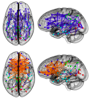
men are map readers and women are intuitive, but bloggers are fast

Connection-wise analysis for males and females (Ingalhalikar et al., 2013).


Blink and you've missed it! Is the news cycle over already? I've been too busy real-working under my rock.
The hardwired difference between male and female brains could explain why men are 'better at map reading'

A pioneering study has shown for the first time that the brains of men and women are wired up differently which could explain some of the stereotypical differences in male and female behaviour, scientists have said.

Researchers found that many of the connections in a typical male brain run between the front and the back of the same side of the brain, whereas in women the connections are more likely to run from side to side between the left and right hemispheres of the brain.
. . .

“In women most of the connections go between left and right across the two hemispheres while in men most of the connections go between the front and the back of the brain,” [Ragini Verma] said.

Because the female connections link the left hemisphere, which is associated with logical thinking, with the right, which is linked with intuition, this could help to explain why women tend to do better than men at intuitive tasks, she added.

“Intuition is thinking without thinking. It's what people call gut feelings. Women tend to be better than men at these kinds of skill which are linked with being good mothers,” Professor Verma said.

Ha, ha, ha! You can't be serious, Professor Verma...

And then we have this gem from the Guardian:
Male and female brains wired differently, scans reveal
. . .

Ragini Verma, a researcher at the University of Pennsylvania, said the greatest surprise was how much the findings supported old stereotypes, with men's brains apparently wired more for perception and co-ordinated actions, and women's for social skills and memory, making them better equipped for multitasking.

"If you look at functional studies, the left of the brain is more for logical thinking, the right of the brain is for more intuitive thinking. So if there's a task that involves doing both of those things, it would seem that women are hardwired to do those better," Verma said. "Women are better at intuitive thinking. Women are better at remembering things. When you talk, women are more emotionally involved – they will listen more."

She added: "I was surprised that it matched a lot of the stereotypes that we think we have in our heads. If I wanted to go to a chef or a hairstylist, they are mainly men." [NOTE: the study population ranged in age from 8 to 22, and I don't think sexual orientation was reported... if we're going to talk stereotypes.]

Within one day of the paper's publication in PNAS (Ingalhalikar et al., 2013), critical blog posts were streaming in to counter the gender stereotypes spouted by the authors themselves. It's the mad new world of rapid-fire post-publication peer review! Trial by Twitter and blog and PubPeer.

I almost feel sorry for the authors, like they've been living in rosy days of yore and weren't aware of the looming backlash, not only to their soundbytes, but to their science.

Although I wasn't able to write a proper post about the paper myself, I suppose I should feel proud that such a community of critics sprang into action on such short notice. PNAS has certainly been target of mine for oh, almost 8 years now...


Links:

asking questions about men and women by looking at teenagers

A quick moan about ‘male’ and ‘female’ brains

Are men better wired to read maps or is it a tired cliché?

New insights into gendered brain wiring, or a perfect case study in neurosexism?


How social media is transforming scientific debate, on Storify.

Men, Women, and Big PNAS Papers

Getting in a Tangle Over Men’s and Women’s Brain Wiring

What's For Breakfast? Fried Girl and Boy Brainz! How Men's And Women's Brains are Dramatically Different And What It All Means.

Discussion at PubPeer


ADDENDUM (Dec 5 2013, 9:45PM)

Brain scans prove there is no difference between male and female brains

We don't have to "wire" our children's brains to reinforce gender stereotypes

So my mushy head is 'hardwired' for girly things, is it? If this is science, I am Richard Dawkins

Extra, Extra! Scientists misunderstand own research!

Study: Male, Female Brains Wired Differently

About that PNAS Article: Journalism and Neurosexism
[Figure 1] is a fascinating example of a series of ontological, technological, and statistical translations leading to a 'wiring diagram', i.e. an ostensibly metaphoric image standing in for a series of evidently absent but detectible and determinative differences. These differences in wiring are then cast into normative social frames and categories. ...

...Now I could be mistaken here - the paper is very dense. But to me, the authors appear to have imagined a Platonic ideal brain connectome that is uni-sexed. One has to ask then: could there be 95 different regions of interest that show the brains are more alike then we thought - probabilistically that is! If not, then it would seem that their model doesn't really reflect the one with which they began.

Reference

M. Ingalhalikar, A. Smith, D. Parker, T. D. Satterthwaite, M. A. Elliott, K. Ruparel, H. Hakonarson, R. E. Gur, R. C. Gur, R. Verma (2013). Sex differences in the structural connectome of the human brain. PNAS. DOI: 10.1073/pnas.1316909110

Subscribe to Post Comments [Atom]

Friday, November 29, 2013

The Phases of Shopping Addiction


The blight of Black Friday is upon us. What better time to look at a recent paper on compulsive shopping?

Sohn and Choi (2013) adopted a qualitative approach and recruited a small group of Korean housewives with problematic shopping habits via consumer news websites. These nine women ranged in age from 28 to 40. The authors identified their target group as individuals with compulsive buying disorder, who reported a "preoccupation with shopping, pre-purchase tension or anxiety, and sense of relief following the purchase defined by Faber and O’Guinn (1992)." The participants all had high scores on the Faber and O’Guinn 14 item "compulsive buying checklist."

The authors conducted in-depth 2 hour interviews with each participant and analyzed the data according to a six-step contents analysis (Kim et al. 1999) that derived concept clusters, subcategories, and categories. Of note are these Five Sequential Phases of Shopping Addiction (Sohn & Choi, 2013):

Phase 1. Retail therapy,Filling up emptiness with shopping”
Phase 2. Denial,Ignoring overconsumption”
Phase 3. Debt-ridden,Ran out of money, while nothing left”
Phase 4. Impulsive buying,Driving ones-self to hasty buying”
Phase 5. Compulsive buying,It is crazy but I cannot stop”

Accompanying these phases are 5 themes, 15 subthemes, and 43 codes (shown in detail below).

- click on image for a larger view -


Do you have a strong urge to purchase the latest Xbox One or PS4 before they sell out? Would you feel anxious if you didn't get one? If so, then you may be in Phase IV, Impulsive Buying.


But if you go to the mall every morning and shop online every day, it's all over. You've reached Phase V, Compulsive Buying.


Reference

Sang-Hee Sohn and Yun-Jung Choi (2013). Phases of Shopping Addiction Evidenced by Experiences of Compulsive Buyers. International Journal of Mental Health and Addiction DOI: 10.1007/s11469-013-9449-y

Subscribe to Post Comments [Atom]

Monday, November 25, 2013

Confessions of a Prosocial Psychopath


Many neuroscientists have been howling about the media coverage surrounding a new book written by UC Irvine Professor Emeritus, Dr. James H. Fallon. This is because unbeknownst to himself for 58 years (or apparently to anyone else, for that matter), he was secretly a psychopath. How did he finally discover this? Did he complete the Psychopathy Checklist and score over 30?

No.

Instead, he diagnosed himself as a psychopath on the basis of his PET scan.


Compared to a control brain (top), neuroscientist James Fallon’s brain (bottom) shows significantly decreased activity in areas of the frontal lobe linked to empathy and morality—anatomical patterns that have been linked with psychopathic behavior. Image via James Fallon in The Smithsonian.


This is a case of reverse inference, assuming that a certain pattern of brain activity indicates a particular behavioral state (or in this case, a specific psychiatric diagnosis). While it may be true at the group level that activity in the ventromedial prefrontal cortex is decreased in psychopaths, it's not possible to diagnose an individual on this basis (at least not with our current state of knowledge).

In fact, Fallon himself initially doubted the ugly possibility that he's lacking in empathy, morality and self-control:
In [his book], Fallon seeks to reconcile how he—a happily married family man—could demonstrate the same anatomical patterns that marked the minds of serial killers.

“I’ve never killed anybody, or raped anyone,” he says. “So the first thing I thought was that maybe my hypothesis was wrong, and that these brain areas are not reflective of psychopathy or murderous behavior.”

Not with certainty, they're not. The Smithsonian article continues:
Eventually, based on further neurological and behavioral research into psychopathy, he decided he was indeed a psychopath—just a relatively good kind, what he and others call a “pro-social psychopath,” someone who has difficulty feeling true empathy for others but still keeps his behavior roughly within socially-acceptable bounds.

A "prosocial psychopath"? Isn't this an oxymoron? Aren't psychopaths antisocial by definition? Eventually, he came to doubt the classification scheme. Psychopathy doesn't appear in the DSM...
...in part because it encompasses such a wide range of symptoms. Not all psychopaths kill; some, like Fallon, exhibit other sorts of psychopathic behavior.

“I’m obnoxiously competitive. I won’t let my grandchildren win games. I’m kind of an asshole, and I do jerky things that piss people off,” he says. “But while I’m aggressive, but my aggression is sublimated. I’d rather beat someone in an argument than beat them up.”

This is psychopathy??

(Perhaps) in response to a snarky query on Twitter ("There goes Hare's PCL" [to paraphrase]), Dr. Fallon noted:


But he looks like such a genial fellow!


Taking a quick look inside The Psychopath Inside (p. 26), we learn that Fallon viewed himself as a "nice, regular guy" who was popular and able to form close friendships with women.1 Should we rely on his brain scan, or on his behavior, when applying such a stigmatizing label?


Confessions of an Extremely Antisocial Female Psychopath

Another book published this year would make an interesting pairing with Fallon's personal discovery neuroscience / memoir: Confessions of a Sociopath, by the anonymous "M. E. Thomas". If you want narcissistic antisocial [but nonviolent] braggadocio, this is the book for you.2 For starters, the author is female, placing her in the minority of those with antisocial personality disorder. She's also a Mormon law professor in California.


"Ms. Thomas" wears the stigmatized behaviors as a badge of pride, although she erroneously calls herself a "sociopath" (which doesn't exist as a diagnostic label). But what do I know? She's SO brilliant – she passed the bar exam without even studying (while everyone else was crying):
I loved getting high marks in school; it meant I could get away with things other students couldn't. When I was young, what thrilled me was the risk of figuring out just how little I could study and still pull off the A. It was the same for being an attorney. During the California bar exam, people were crying from the stress. The convention center where the exam took place looked like a disaster relief center; people made desperate attempts to recall everything they had memorized over the prior eight weeks—weeks that I spent vacationing in Mexico. Despite being woefully ill-prepared by many standards, I was able to maintain calm and focus enough to maximize the knowledge I did have. I passed while others failed.

And she's oh so charming!
You would like me if you met me. I have the kind of smile that is common among television show characters and rare in real life, perfect in its sparkly teeth dimensions and ability to express pleasant invitation. I'm the sort of date you would love to take to your ex's wedding—fun, exciting, the perfect office escort. And I'm just the right amount of successful so that your parents would be thrilled if you brought me home.

But the best take on the book comes from Patrick Bateman, who reviews it for Slate:
I take the elevator up to my apartment and wash my hands and sit in my cream leather chair and chase an Adderall with a J&B and read the book in one sitting. It begins with a psychological evaluation that describes M.E. Thomas as a “prototypical psychopathic personality” manifesting “a ruthless and calculating attitude toward social and interpersonal relationships, and a relative immunity to experiencing negative emotions.” ...
. . .

...She’s into “the fine art of ruining people,” according to the title of Chapter 7. She seduces with charisma, and she cunningly covers her hollowness with superficial charm. She’s a “Nietzschean machine.”

And she violates social norms like it’s her job. Emotionally she takes no prisoners: The high school teacher she falsely accuses of harassment, the friends whose boyfriends she sleeps with just because she can, the colleagues she mind-fucks—they’re all just roadkill...

I'd much rather read Fallon's memoir. He sounds like an upstanding and sympathetic guy, despite the fact that he's related to Lizzy Borden.


Further Reading

The Disconnection of Psychopaths

Born This Way?

I Feel Your Pain... and I Enjoy It

Can Brain Activity Predict Criminal Reoffending?

Are Cognitive Factors Related to Criminal Reoffending?

"None of us are saints"

The Stylized Neuroscience of Psychopaths


Footnotes

1 Interestingly, on page 28 we learn that he developed OCD in junior high, in particular an obsession with Catholicism and morality.

2 Confessions of a Glib Blogger: I haven't read either book.

Subscribe to Post Comments [Atom]

Thursday, November 21, 2013

New Deep Brain Stimulation System Measures Neurotransmitter Release


Wireless Instantaneous Neurotransmitter Concentration Sensing System (WINCS) Patient Module printed circuit board & sterilizable case. (Fig. 1, Kimble et al. 2009).


Last month, the New York Times reported that the Defense Advanced Research Projects Agency (DARPA) will spend $70 million to further the development of technologies that use deep brain stimulation (DBS), which has been highly successful in treating Parkinson's Disease (PD). The SUBNETS program (Systems-Based Neurotechnology for Emerging Therapies) is part of the BRAIN Initiative that aims to "revolutionize our understanding of the human mind."

DARPA issued their call for proposals on October 25. My original take was that the goals were overly ambitious and nearly impossible to achieve within the specified time frame:
To elaborate, over a 5 year period, the successful applicants must conduct clinical trials in human patients with 7 specified psychiatric and neurological disorders (not including PD), some of which have never been treated with DBS. The successful teams will use devices that both stimulate and record neural activity, and provide real-time data that can be decoded as reflecting a particular behavioral state... basically, a futuristic implant that can adjust its own stimulation parameters based on how the patient is doing. At least, that's how I interpret it. 

How close are we to seeing a DBS implant that not only stimulates neural tissue, but also records electrical or chemical signals and then uses this information to adjust the stimulation parameters? Closer than I originally suspected. A recent Nature News article reported on the Mayo Clinic's efforts to develop a DARPAesque, state-of-the-art implant that aims to track brain signals in real time:
Researchers hope that the device will identify the electrical and chemical signals in the brain that correlate in real time with the presence and severity of symptoms, including the tremors experienced by people with Parkinson’s disease. This information could help to uncover where and how DBS exerts its therapeutic effects on the brain, and why it sometimes fails, says Kendall Lee, a neurosurgeon at the Mayo Clinic in Rochester, Minnesota, who is leading the project.
. . .

...Using a method called fast-scan cyclic voltammetry, the device applies a localized voltage change in the brain. This transiently pulls electrons off certain neurotransmitters — the brain chemicals that activate or inhibit neurons — giving rise to electrical currents that can be measured. Each neurotransmitter molecule produces a different electrochemical signature, which can be used to identify it and estimate its concentration every 10 milliseconds.

Studies in awake behaving rats have used fast-scan cyclic voltammetry to measure phasic dopamine release associated with burst firing (Robinson et al., 2003).


Fig, 3 (Robinson et al., 2003). Heterogeneity of electrically evoked dopamine release in the nucleus accumbens of a freely moving rat.


Further information about the device is provided in this article from the Mayo Clinic, which indicates that the WINCS has already been tested in 15 human patients with Parkinson's disease or essential tremor. The study registered in clinicaltrials.gov is described as an Efficacy Study whose primary purpose is basic science:
Neurotransmitter Measurements Using Wireless Instantaneous Neurotransmitter Concentration System (WINCS) During Deep Brain Stimulation Neurosurgery

In this study, the investigators will monitor extracellular neurotransmitter levels using a probe that is able to perform real time electrochemical detection during deep brain stimulation surgery. The overall question this study is designed to answer is: Are there neurotransmitters released during deep brain stimulation?

Interestingly, the primary outcome measure is adenosine1 release recorded by WINCS, and the secondary outcome measure is dopamine release (pre-, during, and post-DBS, over a time frame of 30 min). Adenosine A2A antagonists may extend the duration of action of L-dopa, a primary treatment for PD. Preliminary studies in rats were able to detect subsecond dopamine and adenosine release at an implanted sensor in the striatum during high-frequency stimulation of ascending fibers (Kimble et al., 2009). It seems the early results in patients were also successful in measuring neurotransmitter release.

The WINCS will be integrated with another device, the MINCS (Mayo Investigational Neuromodulation Control System), which is optically linked to WINCS. The entire system is being tested in animal models to deliver brain stimulation wirelessly.

Fig 1B (Chang et al., 2013). Photograph of the MINCS-WINCS hardware showing relative size, optical connection, and recording and stimulating electrode leads. ADC = analog-to-digital converter; DAC = digital-to-analog converter; LPF = low-pass filter; MC = microcontroller; TIA = transimpedance amplifier; V/I Sense = voltage/current sense. Numbers 1 and 4 indicate the microcontrollers; 2 and 3 are the Bluetooth modules.


These developments in DBS devices for Parkinson's disease are very impressive indeed, but DARPA wants to go 7 steps further by developing similar closed-loop systems for use in Post-Traumatic Stress Disorder (PTSD), Major Depression, Borderline Personality Disorder (BPD), General Anxiety Disorder (GAD), Traumatic Brain Injury (TBI), Substance Abuse/Addiction, and Fibromyalgia/Chronic Pain. As I said previously:
To the best of my knowledge, there is no published literature on DBS for PTSD, BPD, GAD [as opposed to OCD], or TBI [except for minimally conscious state]. At clinicaltrials.gov, a Pilot Study of DBS of the Amygdala for Treatment-Refractory Combat PTSD was withdrawn prior to enrollment. There's one DBS trial for TBI that aims to enroll 5 patients over a 4 year period. I couldn't find anything for BPD or GAD (although these disorders might possibly be comorbid in some patients treated for depression or OCD).

Although there is a tremendous amount yet to learn about Parkinson's, much more is known about the pathophysiology of PD than of any of the disorders listed above. But given the list of speakers at a recent Society for Neuroscience symposium on the Mechanisms of Deep Brain Stimulation Efficacy in Neuropsychiatric Disorders, scientists and clinicians at Mayo, Emory, Butler/Brown, and Case Western are certainly working on these problems. $70 million from DARPA would sure come in handy, even if the ambitious endpoints are unattainable within 5 years.


Footnote

1 Caffeine is an adenosine antagonist, but the drugs used as adjunct therapies in PD are istradefylline and preladenant.


References

Chang SY, Kimble CJ, Kim I, Paek SB, Kressin KR, Boesche JB, Whitlock SV, Eaker DR, Kasasbeh A, Horne AE, Blaha CD, Bennet KE, & Lee KH (2013). Development of the Mayo Investigational Neuromodulation Control System: toward a closed-loop electrochemical feedback system for deep brain stimulation. Journal of Neurosurgery. PMID: 24116724

Kimble CJ, Johnson DM, Winter BA, Whitlock SV, Kressin KR, Horne AE, Robinson JC, Bledsoe JM, Tye SJ, Chang SY, Agnesi F, Griessenauer CJ, Covey D, Shon YM, Bennet KE, Garris PA, & Lee KH (2009). Wireless Instantaneous Neurotransmitter Concentration Sensing System (WINCS) for intraoperative neurochemical monitoring. Conference proceedings: Annual International Conference of the IEEE Engineering in Medicine and Biology Society, 2009, 4856-9. PMID: 19963865

Robinson DL, Venton BJ, Heien ML, Wightman RM. (2003). Detecting subsecond dopamine release with fast-scan cyclic voltammetry in vivo. Clin Chem. 49:1763-73.

Nature News link via @GholsonLyon

Subscribe to Post Comments [Atom]

eXTReMe Tracker