Monday, August 13, 2012

The Dark Side of Diagnosis by Brain Scan


Daniel Amen: Pioneer or profiteer?: Psychiatrist Daniel Amen uses brain scans to diagnose mental illness. Most peers say that’s bonkers.


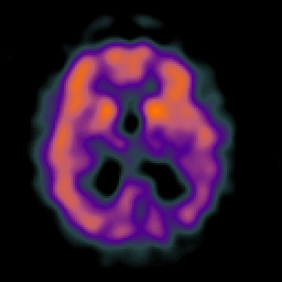
Right on the heels of a Molecular Psychiatry paper that asked, "Why has it taken so long for biological psychiatry to develop clinical tests and what to do about it?" (Kapur et al., 2012) comes this provocatively titled article in the Washington Post about neurohuckster Dr. Daniel Amen and his miraculous SPECT scans:
Daniel Amen is the most popular psychiatrist in America. To most researchers and scientists, that’s a very bad thing.

By , Published: August 9.

NEWPORT BEACH, Calif. — Daniel Amen is, by almost any measure, the most popular psychiatrist in America.

. . .

He has arisen, like a modern-day American myth, from the fields northeast of San Francisco, where he ran a small-town clinic, to become the creator, chairman and CEO of the Amen Clinics, an empire that includes a string of psychiatric practices, a line of nutritional supplements, book publishing, DVD sales, and television and speaking engagements.

. . .

Amen’s career is very troubling, for one of two things must be true.

One, Daniel Gregory Amen, born in 1954 in Encino, Calif., son of Lebanese immigrants, is 20 years ahead of virtually the entire psychiatric field (he says about three dozen other clinics use SPECT scans, but few as profusely as he does), and the establishment has failed to recognize a historic breakthrough.

Or, two, the man has grown fabulously wealthy — he lives in a $4.8 million mansion overlooking the Pacific Ocean — by selling patients a high-priced service that has little scientific validity, yet no regulatory body has made a move to stop him.

SPECT (single photon emission computed tomography) is a relatively inexpensive cousin of PET scanning (positron emission tomography) with lower spatial resolution.1 There is no peer reviewed literature that establishes SPECT as a reliable method of diagnosing psychiatric disorders.

Amen is well-known to regular PBS viewers, because his informercial "Change Your Brain, Change Your Life" [and others] is on regular rotation during fund raisers.2 In a critical Salon.com piece by Robert Burton, one neuroimaging expert was quoted as saying:
"SPECT scans are not sufficiently sensitive or specific to be useful in the diagnosis of A.D.," neurologist Michael Greicius , who runs the Stanford University memory clinic, and has a special interest in the use of functional brain imaging in the diagnosis of A.D., tells me. "The PBS airing of Amen's program provides a stamp of scientific validity to work which has no scientific validity."

In his Washington Post article, author Neely Tucker assembled an impressive list of naysayers:
No major research institution takes his SPECT work seriously, none regards him as “the number one neuroscience guy,” and his revelations, which he presents to rapt audiences as dispatches from the front ranks of science, make the top tier of scientists roll their eyes or get very angry.

“In my opinion, what he’s doing is the modern equivalent of phrenology,” says Jeffrey Lieberman, APA president-elect, author of the textbook “Psychiatry” and chairman of Psychiatry at Columbia University College of Physicians and Surgeons. (Phrenology was the pseudoscience, popular in the early 19th century, that said the mind was determined by the shape of the skull, particularly its bumps.) “The claims he makes are not supported by reliable science, and one has to be skeptical about his motivation.”

“I think you have a vulnerable patient population that doesn’t know any better,” says M. Elizabeth Oates, chair of the Commission on Nuclear Medicine, Board of Chancellors at the American College of Radiology, and chair of the department of radiology at the University of Kentucky.

“A sham,” says Martha J. Farah, director of the Center for Neuroscience & Society at the University of Pennsylvania, summing up her thoughts on one of Amen’s most recent scientific papers.

“I guess we’re all amateurs except for him,” says Helen Mayberg, a psychiatry, neurology and radiology professor at Emory School of Medicine and one of the most respected researchers into depression and brain scanning. “He’s making claims that are outrageous and not supported by any research.”

“I can’t imagine clinical decisions being guided by an imaging test,” says Steven E. Hyman, former director of the National Institute of Mental Health and current director of the Stanley Center for Psychiatric Research at the Broad Institute of MIT and Harvard.

But wait! Didn't The Neurocritic just write a post that expressed some degree of optimism about the prospect of a "stratified psychiatry" of phenotypic or genotypic subtypes at some unspecified future date?

But beware! This is not the same thing as using a SPECT scan to devise a treatment plan. In his NIMH Director's Blog, Dr. Thomas Insel singles out Amen and warns, Brain Scans – Not Quite Ready for Prime Time.


Where Are the Clinical Tests for Psychiatric Disorders?

In their paper, Kapur, Phillips, and Insel (2012) were careful to distinguish their projections for the future from diagnostic tests:3
The prospects of ‘diagnostic tests’ for DSM entities remain distant for reasons articulated above, and it seems unlikely that we will replace the 300-disorder taxonomy of the DSM-5 with an alternative biologically based classification system anytime soon. Therefore the real opportunity for psychiatry is to use the emerging advances in genetics, molecular biology, imaging and cognitive science to supplement, rather than replace, the symptom-driven diagnosis.

Dr. Amen is not so circumspect, in fact he's rather bullish:
He says he has taught himself — by scanning 45,000 people a total of 70,000 times — to apply SPECT, alongside clinical evaluations, as a diagnostic tool in 90 percent of his patients.

The brain activity he says he sees in these scans — areas of high and low activity — allows him to target those areas with specific treatments and medication, he says. A full initial session, including two scans, costs about $3,500.

Amen says this method has helped him identify new subtypes of anxiety, depression and attention deficit disorder, categories far more specific than even the forthcoming fifth edition of the Diagnostic and Statistical Manual of Mental Disorders, the benchmark of the field.

This is heady stuff — using brain imaging to find biomarkers for mental illnesses has been the great hope of psychiatry for at least two decades...


And what are those treatments and medications? Exercise, healthy diet, supplements from his product line [e.g., Serotonin Mood Support], and prescription refills in some cases.


"Due to overwhelming popularity we are now offering the Brain on Joy Bar by the case (18 bars)."


Am I blue, Dr. Amen?

The SPECT scans often seem to be used as either a scare tactic or a form of reassurance:

Amen’s first scan patient, back in the early 1990s, was Sandy.

She was 40, had ADD and had tried to kill herself the night before their initial meeting. In his telling — it is his Genesis story, and he has told it many times — the resulting scan showed a precipitous drop in activity in her prefrontal cortex, the brain’s decision-making center, when she tried to concentrate.

When he showed her the scan, she wept. “You mean it’s not my fault?”

Realizing it was a “biological, not moral” problem, she accepted the diagnosis, took her medications and was greatly helped.

“I thought, ‘Whoa. Pictures matter. You get great compliance,’ ” Amen says.
Another patient said, “The results [of the scan] were a little disconcerting, but I’m glad to have it.”

And here's Amen's colleague Earl Henslin: “If at all possible, I’m motivating my patients to get that scan at the first session. They see that scan and they’re willing to take responsibility.”


A Picture Is Worth a Thousand Dollars

That's the title of a 2009 editorial by Dr. Martha Farah in the Journal of Cognitive Neuroscience. She argues that cognitive neuroscientists have a responsibility to speak out when clinical [and legal] applications of brain imaging are being misrepresented by for-profit companies such as the Amen Clinics:
Tens of thousands of individuals, many of them children, have been exposed to the radiation of two SPECT scans and paid thousands of dollars out of pocket (because insurers will not pay) against the advice of many experts... The Amen Clinics are now marketing their services outside the medical arena, advising couples with marital problems and even “prescreening” couples.

People are swayed by colorful brain images, whether they're in the classroom, the courtroom, or the clinic. Or on reality TV. SPECT scans should not be used for diagnostic or entertainment purposes; there's no scientific evidence for the former and the latter is unethical.


Further Reading

This Is the Presidential Candidates' Brains On...

On Amen's Dec. 2007 editorial in the Los Angeles Times advocating SPECT scans for presidential candidates.

Celebrity Neurostigma
On celebrity SPECT scans from celebrity patients in the reality show Celebrity Rehab with Dr. Drew. Also a lesson in medical ethics. Special guest appearance by Dr. Amen.

Dennis Rodman-Mindy McCready Mind Meld
On the startling similarities in the SPECT scans of Celebrity Rehab participants Dennis Rodman and Mindy McCready.


Footnotes

1 In case you're interested in learning more about how the method works, this review chapter (by the Committee on the Mathematics and Physics of Emerging Dynamic Biomedical Imaging, National Research Council) is one place to start.

2 In fact, I can tell when there's a PBS fundraiser because the number of visitors from a search of daniel amen quack increases.

3 However, new research applies machine learning algorithms to neuroimaging data in an attempt to classify patients with neurological and psychiatric disorders (Orrù et al., 2012).


References

Farah MJ. (2009). A picture is worth a thousand dollars. J Cogn Neurosci. 21:623-4.

Kapur S, Phillips AG, & Insel TR (2012). Why has it taken so long for biological psychiatry to develop clinical tests and what to do about it? Molecular psychiatry PMID: 22869033

Orrù G, Pettersson-Yeo W, Marquand AF, Sartori G, Mechelli A. (2012). Using Support Vector Machine to identify imaging biomarkers of neurological and psychiatric disease: a critical review. Neurosci Biobehav Rev. 36:1140-52.

WaPo link via @vaughanbell.

Subscribe to Post Comments [Atom]

Wednesday, May 21, 2008

More Brain Scams

Speaking of brain scamming, Mind Hacks linked to an article in Salon.com about a disreputable informercial unwittingly shown on PBS multiple times:
Brain scam

Why is PBS airing Dr. Daniel Amen's self-produced infomercial for the prevention of Alzheimer's disease?

By Robert Burton

May. 12, 2008 | It's 10 on a Saturday night and on my local PBS station a diminutive middle-aged doctor with a toothy smile and televangelical delivery is facing a rapt studio audience. "I will show you how to make your brain great, including how to prevent Alzheimer's disease," he declares. "And I'm not kidding."

...the doctor, Daniel Amen, is being interviewed by KQED host Greg Sherwood. Sherwood is wildly enthusiastic. After reading Amen's book, "Change Your Brain, Change Your Life," Sherwood says, "The first thing I wanted to do was to get a brain scan." He turns to Amen. "You could start taking care 10 years in advance of ever having a symptom and prevent Alzheimer's disease," he says. "Yes, prevent Alzheimer's disease," Amen chimes in.

Wait a minute. Prevent Alzheimer's disease? Is he kidding? But Sherwood is already holding up Amen's package of DVDs on learning your risk factors for A.D., as well as his book with a section titled "Preventing Alzheimer's." Then, as though offering a landmark insight into a tragic disease -- and encouraging viewers to pledge money to the station -- Sherwood beams and says, "This is the kind of program that you've come to expect from PBS."

If so, that's a shame...
Back in December, The Neurocritic dissected the neurohuckster's editorial in the Los Angeles Times about SPECT-scanning the brains of presidential candidates:
Getting inside their heads ... really inside

Presidential candidates' health is a campaign issue. So what about their brains?

By Daniel G. Amen
December 5, 2007


. . .

Is the brain health of a presidential candidate a fair topic in an election year? Certainly Dick Cheney's heart condition wasn't off-limits in 2000... Should we go so far as to do brain scans? Of candidates for the Oval Office? Some people might consider discussing brain health a ridiculous idea. Not me.

As a neuropsychiatrist and brain-imaging expert, [NOTE: huh, 9 papers in mostly low-profile journals] I want our elected leaders to be some of the "brain healthiest people" in the land. How do you know about the brain health of a presidential candidate unless you look? ...

Three of the last four presidents have shown clear brain pathology. [NOTE: oh really?? we only have evidence for AD in Reagan, as much as we'd like to believe that George W. has brain damage.] President Reagan's Alzheimer's disease was evident during his second term in office. Nonelected people were covering up his forgetfulness and directing the country's business. Few people knew it, but we had a national crisis. Brain studies have been shown to predict Alzheimer's five to nine years before people have their first symptoms. [NOTE: uh...no. Published studies say 2-3 years.]
SPECT (single photon emission computed tomography) is a relatively inexpensive cousin of PET scanning (positron emission tomography) with lower spatial resolution. The Neurocritic is not all that knowledgeable about SPECT as an imaging method, but these authors are (Committee on the Mathematics and Physics of Emerging Dynamic Biomedical Imaging, National Research Council), in case you're interested in learning more.

Back to Robert Burton and Salon.com:
"SPECT scans are not sufficiently sensitive or specific to be useful in the diagnosis of A.D.," neurologist Michael Greicius , who runs the Stanford University memory clinic, and has a special interest in the use of functional brain imaging in the diagnosis of A.D., tells me. "The PBS airing of Amen's program provides a stamp of scientific validity to work which has no scientific validity."

Throughout March and April this year, "Change Your Brain, Change Your Life" aired nearly 1,300 times on PBS stations across the country, reaching more than 75 percent of U.S. television households. ...the nation's public broadcasting system did not vet "Change Your Brain, Change Your Life" for scientific validity. As a result, it broadcast what amounts to an unregulated infomercial for Amen's unproven treatments.
But it gets even worse:
Amen ... has not followed a traditional scientific path. He received a biology degree from Southern California College, a Pentecostal school, now Vanguard University ("We believe The Bible to be the inspired and only infallible and authoritative Word of God"), and earned his medical degree from Oral Roberts University School of Medicine, defunct as of 1989.

"One of the sustaining factors in my work has been my own personal faith," he declared in his 2002 book... "From the first month that I started to order these (SPECT) scans, I felt that they had a special place in science and that I was led by God to pursue this work."

. . .

And yet Amen's sense of calling hasn't led him to undertake the high-quality clinical investigations that would lend scientific credence to his claims...
Not surprisingly, Amen has his own page at Quackwatch. My question is, how did he get all this free publicity from PBS? Burton had a hard time finding an answer.
In trying to divine PBS' role and obligations in airing such an obviously controversial figure as Amen, I got the proverbial runaround...

Did a local PBS station, or PBS headquarters, do proper vetting? Michael Getler, the PBS ombudsman, didn't have an answer for me and forwarded my message to "the top people." I then got a note from Joseph Campbell, PBS vice president of fundraising programming, who said, "PBS is not responsible for the content of those programs obtained from outside sources (other than PBS); it is up to each individual station to decide on the merits of such non-PBS produced programs."
Daniel Amen responds to "Brain scam"


To learn about more reSPECTable uses of SPECT in Alzheimer's Disease, consult The Whole Brain Atlas.


Here is a mid-ventricular slice which demonstrates the commonest finding in functional imaging of Alzheimer's disease. The dark blue regions in the parietal lobes represent areas of decreased blood flow or perfusion. This reduction in blood flow is due in part to the underlying atrophy, in part to the presence of diseased brain, and in part to the functional "disconnection" of this from other brain regions affected by the disease.

Subscribe to Post Comments [Atom]

Saturday, October 23, 2010

Celebrity Neurostigma


Celebrity SPECT scan from rehab patient


Celebrity Rehab is an American TV reality show on VH1 that exploits the addictions of the rich and C- or D-List famous.
“I thought REAL doctors talked to patients in offices behind closed doors.”
-Lindsay Lohan [who reportedly turned down six figures to appear on the show]
Privacy? Confidentiality? Those rights don't apply to the alcoholic and drug-addicted characters who appear on television and other public media outlets as a form of entertainment. How many of you professional psychology and mental health and cog neuro and pharmaceutical types have taken training courses such as the CITI Course in The Protection of Human Research Subjects? All of you?

Medical Ethics do not apply to Dr. Drew, the star and chief physician of the Celebrity Addiction franchise:
In 2009 [Dr. Drew] Pinsky drew criticism from experts for publicly offering professional opinions of celebrities he has never met or personally examined, based on media accounts, and has also drawn the ire of some of those celebrities.
In contradistinction, proper clinicians who make media appearances take great pains to avoid such unethical outbursts. As explained by Dr Petra Boynton:
They tried to make me talk about rehab but I said ‘no, no, no’

Yesterday I had over 25 emails and phone messages from journalists wanting me to comment on the mental state of several celebrities currently in the press with various drug/relationships problems.

And I’ve said no to all of them.

At the risk of sounding like a broken record here’s why psychologists (and other experts working with the media) can’t talk about celebrities.

If we know the celeb in person (for example as their therapist or healthcare provider) we are breaking their confidence if we speak about them in public. If we do not know them personally we’re simply speculating about them if we were to comment.

The same applies to case studies based on people who are not famous.

In Season 3 Episode #6, ('Triggers') Dr. Drew takes former NBA star Dennis Rodman to see our favorite neurohuckster, Dr. Daniel G. Amen, for a SPECT scan. Amen claims he can diagnose all sorts of psychiatric and neurological ailments using SPECT (single photon emission computed tomography)1 procedures performed at his clinics.

Drew Pinsky exacerbates the unprofessional circus-like atmosphere by making all sorts of unfounded dire predictions about the state of Rodman's brain.

Dr. Drew voiceover: It's day 13, and despite nearly 2 weeks of intense treatment, Dennis has rigidly refused to identify as an alcoholic. It's clear to me there's much more going on here. Probably on an organic basis, both in terms of his personality functioning and possibly damage caused by the alcohol itself. I've arranged for Dennis to receive a brain scan to show him objective evidence of what I suspect is going on.

Van arrives at the Amen Clinics in Newport Beach, CA and Rodman is placed in the scanner.


Cue colorful images of Mr. Rodman's brain appearing on the monitor. All very scientific. Then Dr. Drew introduces him to Dr. Amen.

Amen: "So, we did a study called SPECT that looks at how your brain works. And what we see on your scan here, there's some evidence of alcohol damage. When we see this bumpy appearance, I don't like that. I would worry that you could get something like Alzheimer's disease if you don't do a better job of taking care of your brain. Alcoholic dementia is the second most common cause of dementia in the country. The exciting thing is it can be better but without taking good care of it this is going to deteriorate and get worse."

Rodman: "Uh... it doesn't matter. All right." (Gets up and leaves).

Amen made some rather outrageous statements here. Even though he used the qualifying words something like, there is absolutely no evidence that alcoholism causes the amyloid plaques and neurofibrillary tangles of Alzheimer's disease. In fact, the pathologies are produced by entirely different mechanisms (Aho et al., 2009):
In the present study, no statistically significant influence was observed for alcohol consumption on the extent of neuropathological lesions encountered in the three most common degenerative disorders. Our results indicate that alcohol-related dementia differs from VCI [vascular cognitive impairment], AD [Alzheimer's disease], and DLB [dementia with Lewy bodies]; i.e., it has a different etiology and pathogenesis.
One meta-analysis even found that heavy drinkers did not have an increased risk of dementia of any kind,2 and regular drinkers had a reduced risk (Anstey et al., 2009).

Then brain imaging non-expert Dr. Drew narrates...

Dr. Drew voiceover: Dennis's scans were quite dramatic. In addition to there being an unusual pattern of temporal lobe dysfunction, which confirms my feelings about his personality, he also clearly has damage from alcohol.

Dr. Drew (to Amen): "It makes me sad thinking about it... if he doesn't change."

I see...

Where in the temporal lobe did Dr. Drew find the confirmatory evidence of personality disorder? Anterior temporal lobes (semantic memory)? Posterior/inferior temporal regions, such as the fusiform gyrus (high-level vision)? Superior temporal plane (audition)? Region of the left posterior superior temporal gyrus (Wernicke's area for language comprehension)? Area MT/V5 (perception of motion)? The medial temporal lobe memory system? The amygdala and other portions of the limbic system? Yeah, maybe that, but Amen's "bumpy appearance" was located in ventral visual areas.


Dramatic Brain of Dennis Rodman, age 48

(which looks a lot better than this 38 yr old with 17 years of heavy weekend use).


Scientifically Unfounded Claims in Diagnosing and Treating Patients

That's the title of a Letter to the Editor of the American Journal of Psychiatry by Adinoff and Devous (2010). They responded positively to a critical review (Leuchter, 2009) of none other than Daniel Amen's book, Healing the Hardware of the Soul: Enhance Your Brain to Improve Your Work, Love, and Spiritual Life. The letter goes even further in critiquing Amen's methods:
Several years ago, following conversations with Dr. Amen on how to address such concerns, the Brain Imaging Council of the Society of Nuclear Medicine offered Dr. Amen the opportunity to submit his analyses of a blinded set of SPECT scans (to have been prepared by the Brain Imaging Council) to determine how effective his technique is at correctly diagnosing subjects. Although this proposed study could have provided support for his approach, the offer was declined. Nevertheless, for more than two decades, Dr. Amen has persisted in using scientifically unfounded claims to diagnose and treat patients (over 45,000 by his own count).

There are several dangers to patients that can accrue from this approach: 1) patients (including children) are administered a radioactive isotope without sound clinical rationale; 2) patients pursue treatments contingent upon an interpretation of a SPECT image that lacks empirical support; and 3) based on a presumed diagnosis provided by Dr. Amen's clinics, patients are guided toward treatment that may detract them from clinically sound treatments.

Amen published a rebuttal letter, Brain SPECT Imaging in Clinical Practice (Amen, 2010). Here, he portrays himself as an early adopter -- ahead of his time, persecuted by the unenlightened -- and highly ethical:
The Society of Nuclear Medicine has never formally approached me to perform a study. Plus, I would never engage in a charade where I was expected to give a diagnosis from a scan. That is not how imaging is or should be practiced. The notion of Adinoff and Devous that SPECT is dangerous is disingenuous.
Really? An appearance on Celebrity Rehab and a proclamation that Dennis Rodman is in danger of getting "something like Alzheimer's disease" (based on reading his scan) is NOT a charade?


Public Medical Disclosure

Why is that so inappropriate?
The HIPAA Privacy Rule provides federal protections for personal health information held by covered entities and gives patients an array of rights with respect to that information. At the same time, the Privacy Rule is balanced so that it permits the disclosure of personal health information needed for patient care and other important purposes.
Did Rodman understand all the medical procedures that would be performed on him? Did he sign a consent form in advance to allow the release of a stigmatizing diagnosis for public consumption? Did the fine doctors and television producers consider that if they wanted to portray Rodman as brain damaged, such a compromised mental status could potentially limit his capacity to give informed consent?

But it's only entertainment, after all.

So back to the scientifically unfounded claims... Without mentioning Celebrity Rehab and its diagnostic disclosure issues (perhaps he didn't watch this particular episode), the Editor of AJP felt strongly enough to publish his own Note, appended to the letter of Adinoff and Devous, which concludes:
We have published this exchange of letters as part of our responsibility to readers to point out when a procedure may lack sufficient evidence to justify its widespread clinical use.

For more on Amen and his practices, see The Neuroshrink.

ADDED BONUS! Watch the video for Dr. Drew's neuroanatomy lesson: Celebrity Rehab 3: Dennis's Brain Scan and read the follow-up post: Dennis Rodman-Mindy McCready Mind Meld.


Footnotes

1 SPECT (single photon emission computed tomography) is a less expensive cousin of PET scanning (positron emission tomography) with low spatial resolution. In case you're interested in learning more about it as a valid imaging method, read this from the experts (Committee on the Mathematics and Physics of Emerging Dynamic Biomedical Imaging, National Research Council).

2 The authors noted there could have been a sampling bias.

References

Adinoff, B., & Devous, M. (2010). Scientifically Unfounded Claims in Diagnosing and Treating Patients. American Journal of Psychiatry, 167 (5), 598-598 DOI: 10.1176/appi.ajp.2010.10020157

Aho L, Karkola K, Juusela J, Alafuzoff I. (2009). Heavy alcohol consumption and neuropathological lesions: a post-mortem human study. J Neurosci Res. 87:2786-92.

Amen D. (2010). Brain SPECT Imaging in Clinical Practice. Am J Psychiatry 167: 1125.

Anstey KJ, Mack HA, Cherbuin N. (2009). Alcohol consumption as a risk factor for dementia and cognitive decline: meta-analysis of prospective studies. Am J Geriatr Psychiatry 17:542-55.

Leuchter AF (2009): Healing the hardware of the soul: enhance your brain to improve your work, love, and spiritual life, by Daniel Amen (book review). Am J Psychiatry 166:625.

Subscribe to Post Comments [Atom]

Friday, January 22, 2016

This Neuroimaging Method Has 100% Diagnostic Accuracy (or your money back)

doi:10.1371/journal.pone.0129659.g003

Did you know that SPECT imaging can diagnose PTSD with 100% accuracy (Amen et al., 2015)? Not only that, out of a sample of 397 patients from the Amen Clinic in Newport Beach, SPECT was able to distinguish between four different groups with 100% accuracy! That's right, the scans of (1) healthy participants, and patients with (2) classic post-traumatic stress disorder (PTSD), (3) classic traumatic brain injury (TBI), and (4) both disorders..... were all classified with 100% accuracy!

TRACK-TBI investigators, your 3T structural and functional MRI outcome measures are obsolete.

NIMH, the hard work of developing biomarkers for mental illness is done, you can shut down now. Except none of this research was funded by you...

The finding was #19 in a list of the top 100 stories by Discover Magazine.


How could the Amen Clinics, a for-profit commercial enterprise, accomplish what an army of investigators with billions in federal funding could not?

The authors1 relied on a large database of scans collected from multiple sites over a 20 year period. The total sample included 20,746 individuals who visited one of nine Amen Clinics from 1995-2014 for psychiatric and/or neurological evaluation (Amen et al., 2015). The first analysis included a smaller, highly selected sample matched on a number of dimensions, including psychiatric comorbidities (Group 1).

- click on image for larger view -


You'll notice the percentage of patients with ADHD was remarkably high (58%, matched across the three patient groups). Perhaps that's because...


I did not know that.
 Featuring Johnny Cash ADD.


SPECT uses a radioactive tracer injected 30 minutes before a scan that will assess either the “resting state” or an “on-task” condition (a continuous performance task, in this study). Clearly, SPECT is not the go-to method if you're looking for decent temporal resolution to compare two conditions of an active attention task. The authors used a region of interest (ROI) analysis to measure tracer activity (counts) in specific brain regions.

I wondered about the circularity of the clinical diagnosis (i.e., were the SPECT scans used to aid diagnosis), particularly since “Diagnoses were made by board certified or eligible psychiatrists, using all of the data available to them, including detailed clinical history, mental status examination and DSM-IV or V criteria...” But we were assured that wasn't the case: “These quantitative ROI metrics were in no way used to aid in the clinical diagnosis of PTSD or TBI.” The rest of the methods (see Footnote 2) were opaque to me, as I know nothing about SPECT.

A second analysis relied on visual readings (VR) of about 30 cortical and subcortical ROIs. “Raters did not have access to detailed clinical information, but did know age, gender, medications, and primary presenting symptoms (ex. depressive symptoms, apathy, etc.).”  Hmm...

But the quantitative ROI analysis gave superior results to the clinician VR. So superior, in fact, that the sensitivity/specificity in distinguishing one group from another was 100% (indicated by red boxes below). The VR distinguished patients from controls with 100% accuracy, but was not as good for classifying the different patient groups during the resting state scan — only a measly 86% sensitivity, 81% specificity for TBI vs. PTSD, which is still much better than other studies. However, results from the massively sized Group 2 were completely unimpressive. 3


- click on image for larger view, you'll want to see this -


Why is this so important? PTSD and TBI can show overlapping symptoms in war veterans and civilians alike, and the disorders can co-occur in the same individual. More accurate diagnosis can lead to better treatments. This active area of research is nicely reviewed in the paper, but no major breakthroughs have been reported yet. So the claims of Amen et al. are remarkable. Stunning if true. But they're not. They can't be. The accuracy of the classifier exceeds the precision of the measurements, so this can't be possible. What is the test-retest reliability of SPECT? What is the concordance across sites? Was there no change in imaging protocol, no improvements or upgrades to the equipment over 20 years? SPECT is sensitive to motion artifact, so how was that handled, especially in patients who purportedly have ADHD?

SPECT has been noted for its poor spatial resolution compared to other functional neuroimaging techniques like PET and fMRI. A panel of 16 experts did not include SPECT among the recommended imaging modalities for the detection of TBI. Dr. Amen and his Clinics in particular have been criticized in journals (Farah, 2009; Adinoff & Devous, 2010a, 2010b; Chancellor &, Chatterjee, 2011) and blogs (Science-Based Medicine, The Neurocritic, and Neurobollocks) for making unsubstantiated claims about the diagnostic accuracy and usefulness of SPECT.

Are his latest results too good to be true? You can check for yourself! The paper was published in PLOS ONE, which has an open data policy:
PLOS journals require authors to make all data underlying the findings described in their manuscript fully available without restriction, with rare exception.

When submitting a manuscript online, authors must provide a Data Availability Statement describing compliance with PLOS's policy. If the article is accepted for publication, the data availability statement will be published as part of the final article.

Before you get too excited, here's the Data Availability Statement:
Data Availability: All relevant data are within the paper.

But this is not true. NONE of the data are available within the paper. There's no way to reproduce the authors' analyses, or to conduct your own. This is a problem, because...
Refusal to share data and related metadata and methods in accordance with this policy will be grounds for rejection. PLOS journal editors encourage researchers to contact them if they encounter difficulties in obtaining data from articles published in PLOS journals. If restrictions on access to data come to light after publication, we reserve the right to post a correction, to contact the authors' institutions and funders, or in extreme cases to retract the publication.

So all you “research parasites” out there4 — you can request the data. I thought this modest proposal would create a brouhaha until I saw a 2014 press release announcing the World's Largest Database of Functional Brain Scans Produces New Insights to Help Better Diagnose and Treat Mental Health Issues:
With a generous grant from the Seeds Foundation [a Christian philanthropic organization] in Hong Kong, Dr. Amen and his research team led by neuroscientist Kristen Willeumier, PhD, have turned the de-identified scans and clinical information into a searchable database that is shared with other researchers around the world.

In the last two years, Amen and colleagues have presented 20 posters at the National Academy of Neuropsychology. The PR continues:
The magnitude and clinical significance of the Amen Clinics database – being the world's largest SPECT imaging database having such volume and breadth of data from patients 9 months old to 101 years of age – makes it a treasure trove for researchers to help advance and revolutionize the practice of psychiatry.

Does this mean that Dr. Amen will grant you access to the PLOS ONE dataset (or to the entire Amen Clinics database) if you ask nicely? If anyone tries to do this, please leave a comment.


Footnotes

1 The other authors included Dr. Andrew “Glossolalia” Newberg and Dr. Theodore “Neuro-Luminance Synaptic Space” Henderson.

2 Methods:
To account for outliers, T-score derived ROI count measurements were derived using trimmed means [91] that are calculated using all scores within the 98% confidence interval (-2.58 < Z < -2.58). The ROI mean for each subject and the trimmed mean for the sample are used to calculate T with the following formula: T = 10*((subject ROI_mean - trimmed regional_avg)/trimmed regional_stdev)+50.
3 Results from the less pristine Group 2 were not impressive at all, I must say. Group 2 had TBI (n=7,505), PTSD (n=1,077), or both (n=1,017) compared to n=11,147 patients without either (these were not clean controls as in Group 1). Given the massive number of subjects, the results were clinically useless, for the most part (see Table 6).

4 A brand new editorial in NEJM by Longo and Drazen (who decry “research parasites”) is causing a twitterstorm with the hashtags #researchparasites and #IAmAResearchParasite.


References

Adinoff B, Devous M. (2010a). Scientifically unfounded claims in diagnosing and treating patients. Am J Psychiatry 167(5):598.

Adinoff B, Devous M. (2010b). Response to Amen letter. Am J Psychiatry 167(9):1125-1126.

Amen, D., Raji, C., Willeumier, K., Taylor, D., Tarzwell, R., Newberg, A., & Henderson, T. (2015). Functional Neuroimaging Distinguishes Posttraumatic Stress Disorder from Traumatic Brain Injury in Focused and Large Community Datasets PLOS ONE, 10 (7) DOI: 10.1371/journal.pone.0129659

Chancellor B, Chatterjee A. (2011). Brain branding: When neuroscience and commerce collide. AJOB Neuroscience 2(4): 18-27.

Farah MJ. (2009). A picture is worth a thousand dollars. J Cogn Neurosci. 21(4):623-4.

Subscribe to Post Comments [Atom]

Saturday, April 30, 2016

The Truth About Cognitive Impairment in Retired NFL Players


NINETY-TWO percent of retired National Football League players have decreased cognitive function, according to a new study:
“In the NFL group, baseline neuropsychological assessments showed 92% of players had decreased general cognitive proficiency, 86% had decreased information processing speed, 83% had memory loss, 83% had attentional deficits, and 85% had executive function impairment.”

The Truth?

The study reported on a self-selected sample of 161 current and retired NFL players recruited via a blog (“The NFL concealed the danger of brain injuries!!”), the Los Angeles Chapter of the Retired NFL Players Association, The Summit (??), and possibly other sources. Perhaps these players were motivated to participate because they had cognitive complaints, or because they wanted an evaluation in advance of the $1 billion concussion settlement. The League's Baseline Assessment Program is a required part of the settlement.1

The quote above is the full extent of the report on the players' neuropsychological assessments. These were done using computerized test batteries (MicroCog or WebNeuro), which are largely unknown to most clinical neuropsychologists. Was there an adequately matched control population? What norms were used? They don't say.

THE TRUTH IS, we don't know the extent of cognitive impairment in these football players, or the percentage of all players who are affected, or the severity of impairment in those who are. This new paper (by Daniel Amen, Bennet Omalu, and others) doesn't give us enough information, but it succeeds in sounding the alarm about the dangers of football and the inevitability of memory loss and attention deficits.

Are blows to the head bad for your brain? Can repeated concussions cause cognitive impairment and chronic traumatic encephalopathy (CTE)? 2  Yes, almost certainly, but we can't rely on biased samples, appeal to celebrity, and Frontline documentaries (“researchers have identified CTE in 96 percent of NFL players that they’ve examined”) as conclusive scientific evidence. What's needed are better sampling methods (in the short term) and longitudinal studies that follow a diverse cohort over time (in the long term).

The Scans

Caption for top figure: SPECT brain scans showing abnormal low blood flow in an NFL player compared to a normal healthy control subject.

The new paper by Amen et al. (2016) was actually focused on SPECT scans, not surprisingly, since these are the backbone of his business at the Amen Clinics. The article claims “90% sensitivity, 86% specificity, and 94% accuracy” in discriminating NFL players from controls. I won't elaborate here, but check out This Neuroimaging Method Has 100% Diagnostic Accuracy (or your money back) and The Dark Side of Diagnosis by Brain Scan for detailed critiques of the methods used here. I will flag one tiny issue, however:
“All NFL players were male, while 56% of the control group were women.”

Why?? The authors have a database of 100,000 SPECT scans...


Footnote

1  11. What is the Baseline Assessment Program (“BAP”)?
. . .
Retired players who are diagnosed with Level 1 Neurocognitive Impairment (i.e., moderate cognitive impairment) are eligible to receive further medical testing and/or treatment (including counseling and pharmaceuticals) for that condition during the ten-year term of the BAP or within five years from diagnosis, whichever is later.

14. What diagnoses qualify for monetary awards?
Monetary awards are available for the diagnosis of ALS, Parkinson’s Disease, Alzheimer’s Disease, Level 2 Neurocognitive Impairment (i.e., moderate Dementia), Level 1.5 Neurocognitive Impairment (i.e., early Dementia) or Death with CTE (the “Qualifying Diagnoses”). A Qualifying Diagnosis may occur at any time until the end of the 65-year term of the Monetary Award Fund.

2 ADDENDUM (May 1 2016): I should say, “...cause CTE and/or other neurodegenerative disorders and dementias.”

Also see: Here’s What We Don’t Know About Head Injuries And Sport
...and A Clinical Approach to the Diagnosis of Traumatic Encephalopathy Syndrome


Reference

Daniel G. Amen, Kristen Willeumier, Bennet Omalu, Andrew Newberg, Cauligi Raghavendra, & Cyrus A. Raji (2016). Perfusion Neuroimaging Abnormalities Alone Distinguish National Football League Players from a Healthy Population Journal of Alzheimer's Disease : 10.3233/JAD-160207


Caption (from press materials): SPECT brain scans showing improvement of abnormal low blood flow in an NFL player compared after 3.5 months on a customized brain rehabilitation program.

ADDENDUM #2 (May 1 2016): The authors' Conflict of Interest statements.

Subscribe to Post Comments [Atom]

Monday, October 25, 2010

Dennis Rodman-Mindy McCready Mind Meld


Dennis Rodman (left) and Mindy McCready (right) prepare for SPECT scanning at the Amen Clinic in Newport Beach.

Celebrity Rehab with Dr. Drew, part of VH1's Celebreality block of programming, is an American reality show that exploits the addictions of the rich and formerly famous. The previous post, Celebrity Neurostigma, examined the medical ethics (or lack thereof) of practicing physicians who disclose the personal information of patients in their care. Furthermore, Dr. Drew Pinsky regularly offers unsolicited diagnoses of celebrities not in his care, which proved to be embarrassing in the wake of the Joaquin Phoenix hoax.

In Season 3 of the show, Dr. Drew joined forces with self-help author and psychiatrist Dr. Daniel Amen, who was criticized in the American Journal of Psychiatry for his Scientifically Unfounded Claims in Diagnosing and Treating Patients. In his defense, Amen wrote:

"...I would never engage in a charade where I was expected to give a diagnosis from a scan. That is not how imaging is or should be practiced."

-Daniel Amen, M.D. (2010). Brain SPECT Imaging in Clinical Practice. Am J Psychiatry 167: 1125.

Yet, we see him doing just that for two of the celebrity rehab patients, as shown in these short video clips: Dennis's Brain and Mindy's Brain. The images below illustrate their "serious" and "quite dramatic" scans. Rodman is said to have "an unusual pattern of temporal lobe dysfunction" while McCready's temporal lobes "look like they've been hurt."1


This is the Brain of Dennis Rodman, age 48


This is the Brain of Mindy McCready, age 34

Notice any similarities??


ADDENDUM: Obviously, someone is being deceived here. It's either:

(1) The viewers - this is the more acceptable option. Rodman and McCready were actually shown images of their real brains, but judicious off-camera editing replaced this private medical information with stock footage of an archival scan. Or maybe they knew about the farcical aspects and were told to play along as part of the script. OR it's:

(2) The patients - this sort of medical deception, for theatrical "scared straight" purposes, is unethical. Being shown a stranger's brain scan and then being told that it's your damaged temporal lobe, that you're at risk of dementia, that you have alcohol damage, can be considered medical fraud.


Footnote

1 The best part of the clip is when McCready gives a more credible account for the putative abnormalities in her brain: the terrible assault that potentially caused temporal lobe injury.

Subscribe to Post Comments [Atom]

Sunday, April 26, 2015

FDA says no to marketing FDDNP for CTE


The U.S. Food and Drug Administration recently admonished TauMark™, a brain diagnostics company, for advertising brain scans that can diagnose chronic traumatic encephalopathy (CTE), Alzheimer's disease, and other types of dementia. The Los Angeles Times reported that the FDA ordered UCLA researcher Dr. Gary Small and his colleague/business partner Dr. Jorge Barrio to remove misleading information from their company website (example shown below).


CTE has been in the news because the neurodegenerative condition has been linked to a rash of suicides in retired NFL players, based on post-mortem observations. And the TauMark™ group made headlines two years ago with a preliminary study claiming that CTE pathology is detectable in living players (Small et al., 2013).

The FDA letter stated:
The website suggests in a promotional context that FDDNP, an investigational new drug, is safe and effective for the purpose for which it is being investigated or otherwise promotes the drug. As a result, FDDNP is misbranded under section 502(f)(1) of the FD&C Act...

[18F]-FDDNP1 is a molecular imaging probe that crosses the blood brain barrier and binds to several kinds of abnormal proteins in the brain. When tagged with a radioactive tracer, FDDNP can be visualized using PET (positron emission tomography).

Despite what the name of the company implies, FDDNP is not an exclusive tau marker. FDDNP may bind to tau protein [although this is disputed],2 but it also binds to beta-amyloid, found in the clumpy plaques that form in the brains of those with Alzheimer's disease. Tau is found in neurofibrillary tangles, also characteristic of Alzheimer's pathology, and seen in other neurodegenerative tauopathies such as CTE.

The big deal with this and other radiotracers is that the pathological proteins can now be visualized in living human beings. Previously, an Alzheimer's diagnosis could only be given at autopsy, when the post-mortem brain tissue was processed to reveal plaques and tangles. So PET imaging is a BIG improvement. But still, a scan alone is not completely diagnostic, as noted by the Alzheimer's Association:
Even though amyloid plaques in the brain are a characteristic feature of Alzheimer's disease, their presence cannot be used to diagnose the disease. Many people have amyloid plaques in the brain but have no symptoms of cognitive decline or Alzheimer's disease. Because amyloid plaques cannot be used to diagnose Alzheimer's disease, amyloid imaging is not recommended for routine use in patients suspected of having Alzheimer's disease.

from TauMark's old website


There are currently three FDA-approved molecular tracers that bind to beta-amyloid: florbetapir, flutemetamol, and florbetaben (note that none of these is FDDNP). But the big selling point of TauMark™ is (of course) the tau marker part, which would also label tau in the brains of individuals with CTE and frontotemporal dementia, diseases not characterized by amyloid plaques. But how can you tell the difference, when FDDNP targets plaques and tangles (and prion proteins, for that matter)?

A new study by the UCLA team demonstrated that the distribution of FDDNP labeling in the brains of Alzheimer's patients differs from that seen in a selected group of former NFL players with cognitive complaints (Barrio et al., 2015). These retired athletes (and others with a history of multiple concussions) are at risk of developing the brain pathology known as chronic traumatic encephalopathy.


from Fig. 1 (Barrio et al., 2015).  mTBI = mild traumatic brain injury, or concussion. T1 to T4 = progressive FDDNP PET signal patterns.


It's a well-established fact that brains with Alzheimer's disease, frontotemporal lobar degeneration, or Lou Gehrig's disease (for example) all show different patterns of neurodegeneration, so why not extend this to CTE? This may seem like a reasonable approach, but there are problems with some of the assumptions.


Perhaps the most deceptive claim is that “TauMark owns the exclusive license of the first and only brain measure of tau protein...” Au contraire! A review of recent developments in tau PET imaging (Zimmer et al., 2014) said that...
...six novel tau imaging agents—[18F]THK523, [18F]THK5105, [18F]THK5117, [18F]T807, [18F]T808, and [11C]PBB3—have been described and are considered promising as potential tau radioligands.

Note that [18F]FDDNP is not among the six.2,3  In fact, Zimmer et al. (2014) mentioned that in brain slices, “[3H]FDDNP failed to demonstrate overt labeling of tau pathology.” 2

No matter. Former NFL players are clamoring to participate in the TauMark studies.


So to recap, the FDA considered TauMark marketing to be “concerning from a public health perspective.” Their letter warned:
Your website describes FDDNP for use in brain PET scans to diagnose traumatic brain injuries, Alzheimer’s disease, and other neurological conditions. These uses are ones for which a prescription would be needed because they require the supervision of a physician and adequate directions for lay use cannot be written.
(see also Regulatory Focus News and the FDA's own PDF archive of the TauMark site).


At this point, astute followers of The Neurocritic and Neurobollocks might ask, “Hey, how does Dr. Daniel Amen get away with claiming that his SPECT scans can accurately diagnose different types of dementia, each with different ‘treatment plans’?”


Hey FDA, what gives?? Dr. Small and Dr. Barrio have at least 37 peer-reviewed publications on their FDDNP methods and imaging results. Meanwhile, Dr. Amen has two non-peer reviewed poster abstracts on his SPECT results in dementia. With ads like these and appearances on Celebrity Rehab, aren't the Amen Clinics's claims “misbranded” too?


Further Reading

Is CTE Detectable in Living NFL Players?

The Ethics of Public Diagnosis Using an Unvalidated Method

Uncertain Diagnoses, Research Data Privacy, & Preference Heterogeneity

Blast Wave Injury and Chronic Traumatic Encephalopathy: What's the Connection?

Little Evidence for a Direct Link between PTSD and Chronic Traumatic Encephalopathy


Footnotes

1 FDDNP is 2-(1-(6-[(2-[(18)F]fluoroethyl)(methyl)amino]-2-naphthyl)ethylidene)malononitrile.

2 Or what is presumed to be tau. FDDNP is supposedly a tracer for both tau and amyloid, but some experts think it's neither. Zimmer et al. (2014) stated:
Though ... [18F]FDDNP appeared to bind both amyloid plaques and tau tangles, a subsequent study using [3H]FDDNP autoradiography in sections containing neurofibrillary tangles (NFTs) failed to demonstrate overt labeling of tau pathology because of a low affinity for NFTs.
Other studies have shown that it binds to a variety of misfolded proteins.

3 James et al. (2015) were more generous in their review of tau PET imaging, mentioning the existence of seven tau tracers (including FDDNP). But again they noted the lack of specificity.  (Parenthetically speaking, [18F]T807 imaging has been done in a single NFL player, which may be of interest in a future post.)


References

Barrio, J., Small, G., Wong, K., Huang, S., Liu, J., Merrill, D., Giza, C., Fitzsimmons, R., Omalu, B., Bailes, J., & Kepe, V. (2015). In vivo characterization of chronic traumatic encephalopathy using [F-18]FDDNP PET brain imaging. Proceedings of the National Academy of Sciences DOI: 10.1073/pnas.1409952112

Zimmer, E., Leuzy, A., Gauthier, S., & Rosa-Neto, P. (2014). Developments in Tau PET Imaging. The Canadian Journal of Neurological Sciences, 41 (05), 547-553 DOI: 10.1017/cjn.2014.15

Subscribe to Post Comments [Atom]

eXTReMe Tracker